CT -Gastric foreign body, splenic mass and hip dysplasia in a 10 year old M Golden Retriever

Case Study

CT -Gastric foreign body, splenic mass and hip dysplasia in a 10 year old M Golden Retriever

This 10 year old M Golden Retriever presented for lameness left front; rads show possible gastric foreign body, prominent liver, bulbous caudal spleen, spondylosis on rads. 

This 10 year old M Golden Retriever presented for lameness left front; rads show possible gastric foreign body, prominent liver, bulbous caudal spleen, spondylosis on rads. 

DX

Gastric obstruction with obstruction of gastric outflow.Splenic mass lesion.

Image Interpretation

CT of the thorax and abdomen:

Abdomen – 

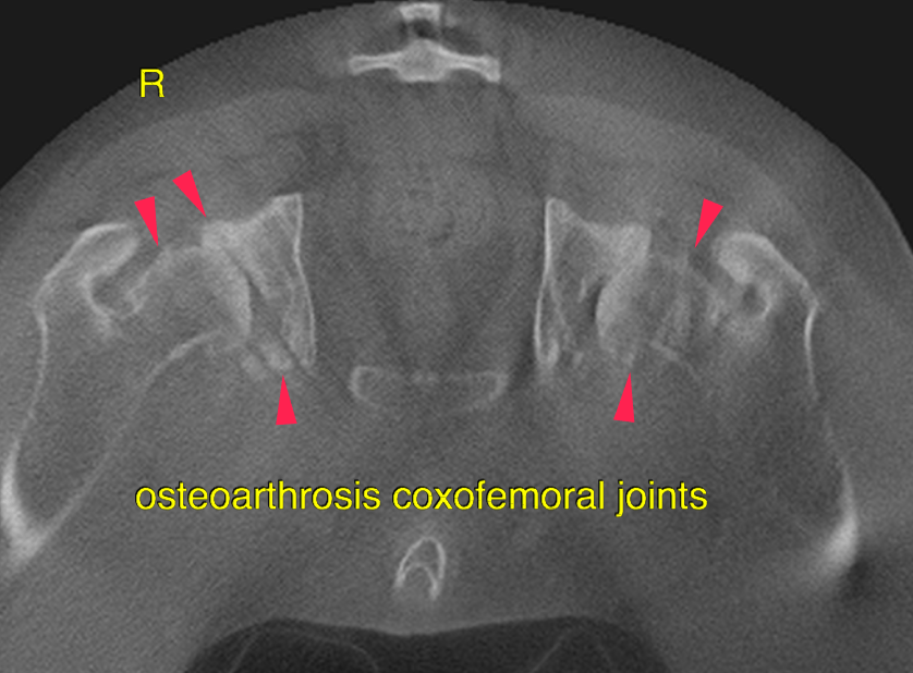
The liver presents with normal shape, even surface, uniformly attenuating parenchyma and homogeneous contrast enhancement, unremarkable.  The tail of the spleen presents a homogeneous contrast enhancing mass lesion, measuring 2.8 cm in diameter. Focal capsular expansion is noted at this level.  Structured soft tissue attenuating material with layered appearance is noted in the gastric fundus and the pyloric antrum. The material within the fundus is bunched up. Between the layered margins of the material, small gas inclusions are present.  At the ventral aspect of multiple vertebral endplates of the lumbar spine spondylosis formation is present. At the level of the intervertebral disc space mild irregular broad based mineral attenuating material is present in the ventral aspect of the vertebral canal L2/L3. It occupies approximately 15% of the diameter of the vertebral canal and the dural tube is mildly displaced dorsally. Both coxofemoral joints present marked osteophyte new bone formation. The femoral heads are dislocated dorsally and the acetabula are shallow. 

Outcome

(socks) and mechanical obstruction. The material obstructs the gastric outflow.
However it did not extend into the descending duodenum at the time of the CT
examination.
The mass lesion in the spleen is consistent with either benign nodular hyperplasia or
primary neoplasia (hemangiosarcoma, roundcell neoplasia).
As the mass lesion in the spleen has a rather solid appearance ultrasound guided FNA
samples of the splenic mass and hypogastric or medial iliac lymph nodes is
recommended to rule out round-cell neoplasia. Splenectomy may be considered
paralleling possible surgical removal of the gastric foreign material alternatively. Osteoarthrosis both coxofemoral joints due to hip-dysplasia. If clinical signs are
present anti-inflammatory drugs are indicated.

Patient Information

Patient Name : Brandon DeFreites/MPI
Gender : Male, Intact
Species : Canine
Type of Imaging : Ultrasound
Status : Complete
Liz Wuz Here : Yes
Code : 16_00136

Clinical Signs

  • Lameness
  • Vomiting

History

  • Foreign Body

Images

bildschirmfoto_2016-09-01_um_11bildschirmfoto_2016-09-01_um_11bildschirmfoto_2016-09-01_um_11bildschirmfoto_2016-09-01_um_11

Clinical Signs

  • Lameness
  • Vomiting
Skip to content