CT – Extradural mass lesion at C6/7 with lateralized disc extrusion in a 11 year old MN Dachshund

Case Study

CT – Extradural mass lesion at C6/7 with lateralized disc extrusion in a 11 year old MN Dachshund

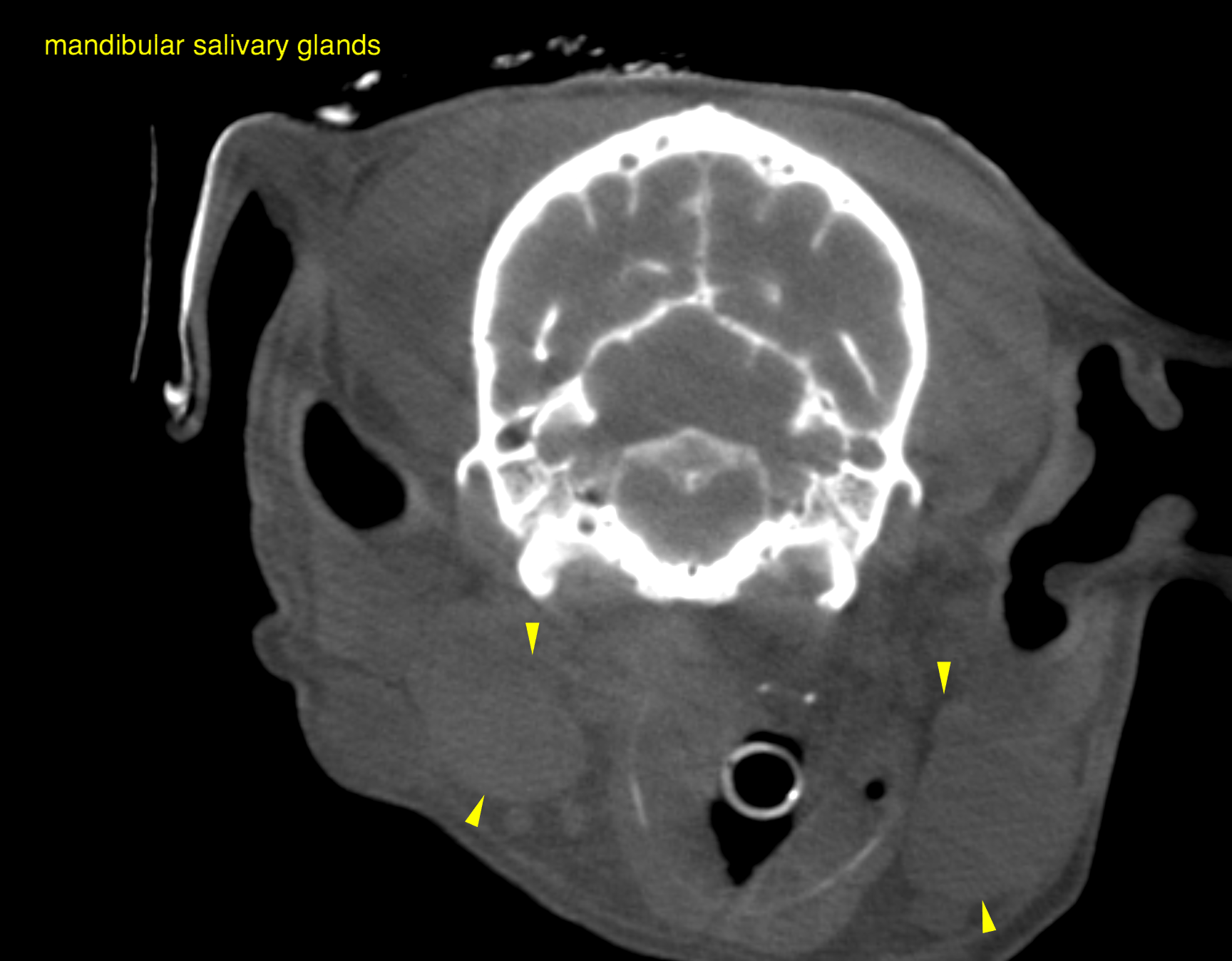
This 11 year old MN Dachshund dog presented 2 weeks prior with inability to close mouth, swollen submandibular lymph nodes or salivary glands and weakness/ataxia. Signs resolved with clindamycin and meloxicam. Patient then presented with left forelimb weakness to left lateralizing tetraparesis.
This 11 year old MN Dachshund dog presented 2 weeks prior with inability to close mouth, swollen submandibular lymph nodes or salivary glands and weakness/ataxia. Signs resolved with clindamycin and meloxicam. Patient then presented with left forelimb weakness to left lateralizing tetraparesis.
DX

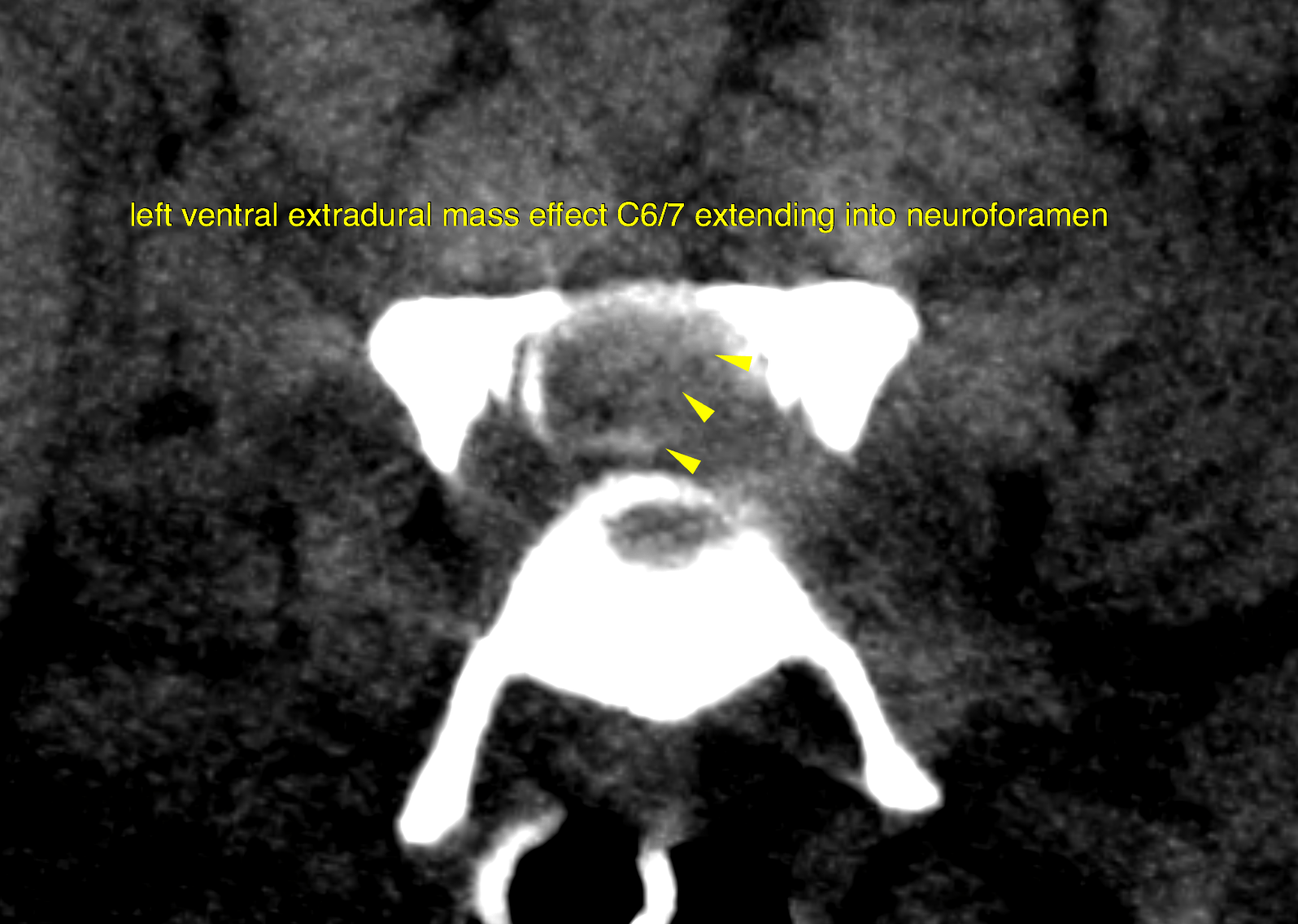
The computed tomography reveals a left-sided and ventral extradural mass lesion with neuroforaminal extension at C6/7.

Image Interpretation

CT of the head and cervical spine – 


 Cervical spine: 

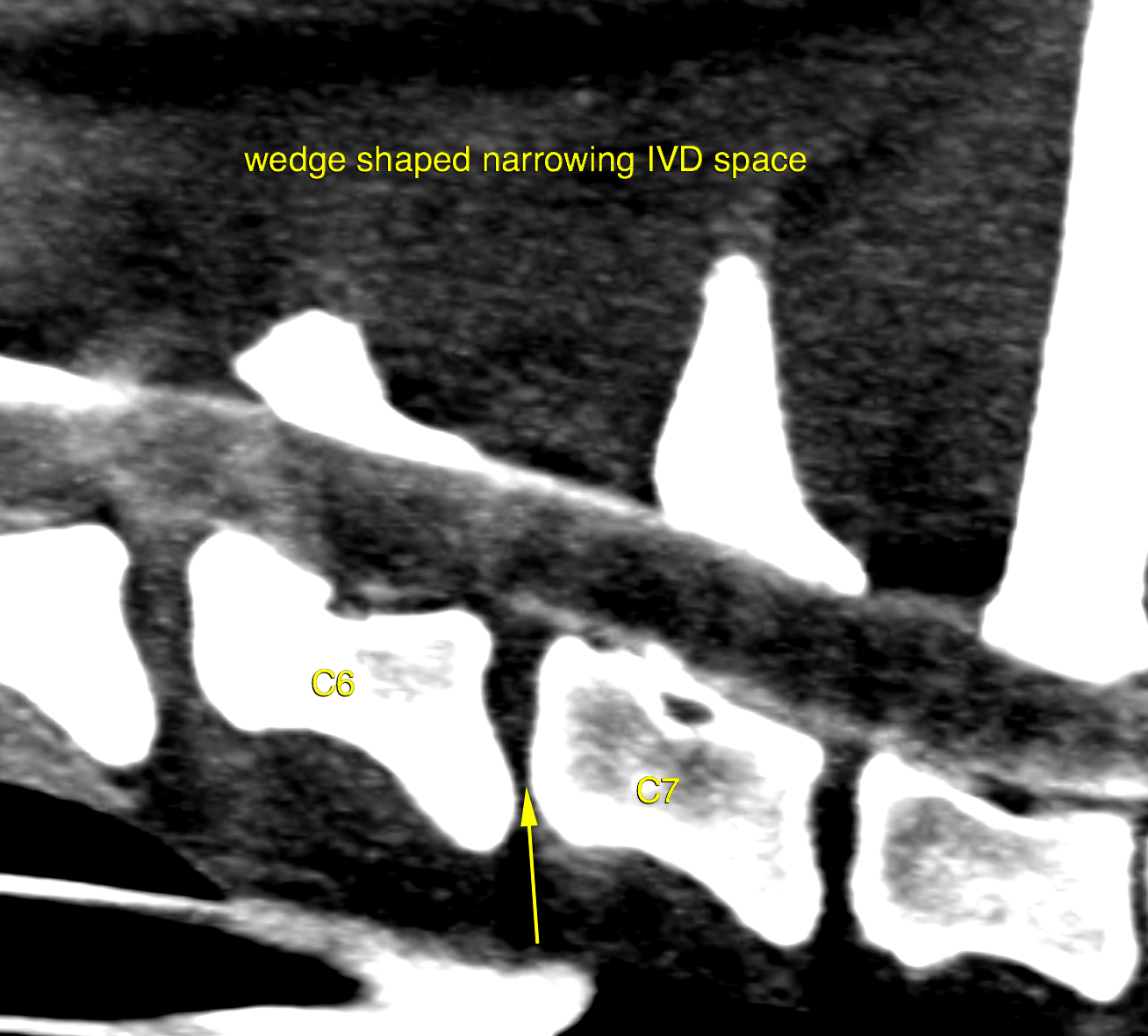
An isoattenuating mass effect with left-sided neuroforaminal extension is noted within the left and ventral epidural space at C6/7. The mass effect displaces and collapses the left and ventral subarachnoid space. The cross sectional area of the vertebral canal is reduced by 40%. The intervertebral disc space reveals ventral wedge shaped narrowing. There is no widening of the neuroforamen. Caudal to the lesion the filling of the subarachnoid space diminishes. Preferential cranial flow of the contrast medium is noted. 

Head: 

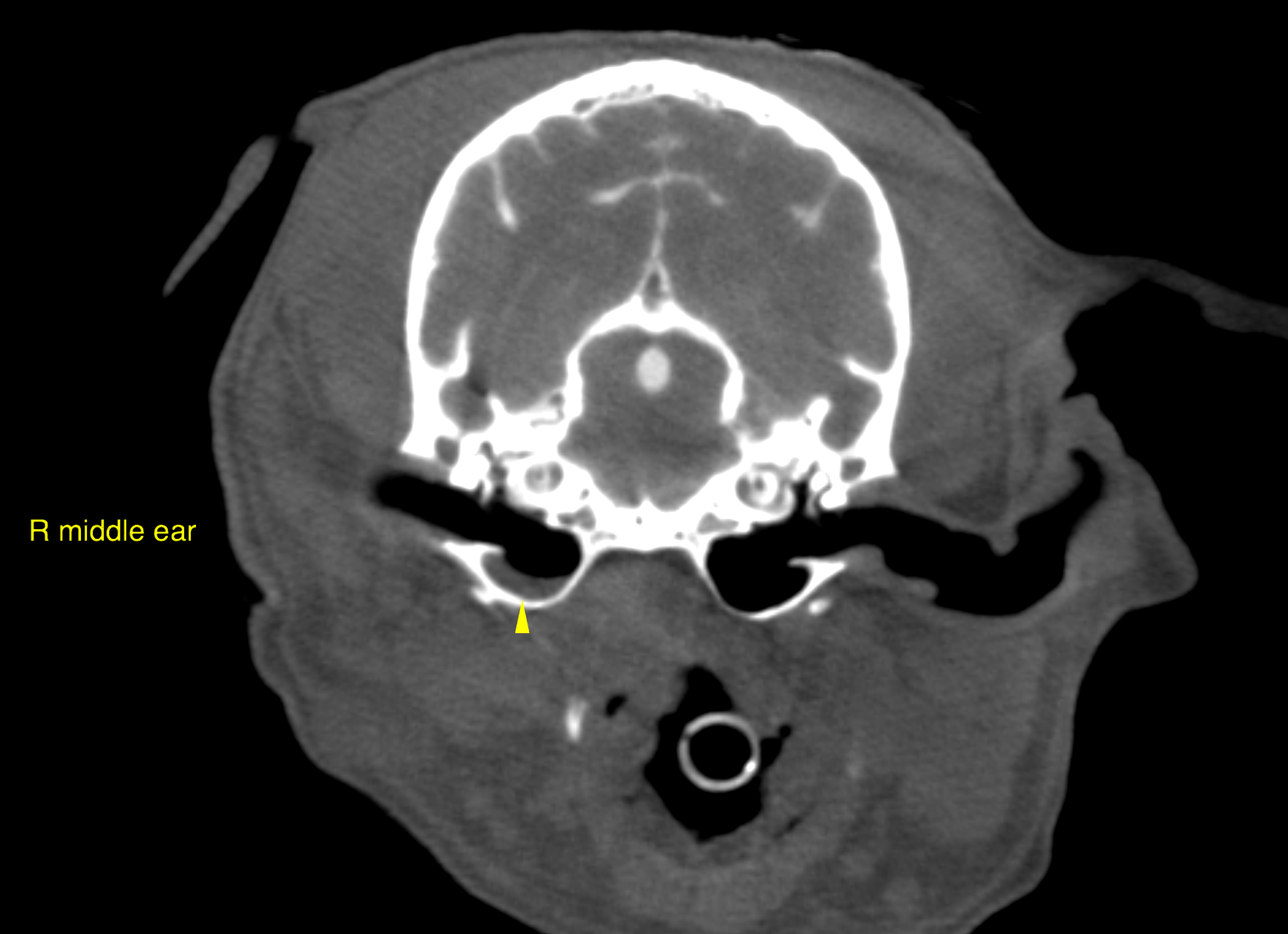
Mild right-sided otitis media is noted by hypoattenuating material with a meniscal surface within the ventral third of the right tympanic bulla. The bulla wall reveals mild and even thickening. 

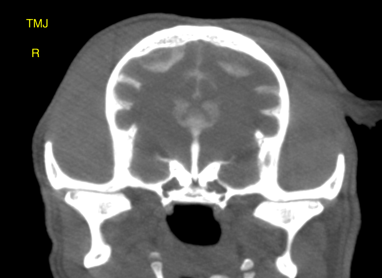
Both temporomandibular joints present mild to moderate signs of a chronic degenerative osteoarthropathy. 

Outcome

An isoattenuating lateralized disc extrusion is very likely and further supported by the narrowing of the respective intervertebral disc space. A peripheral nerve sheath tumor is a low potential here because of the lack of neuroforaminal widening. Surgical exploration with a lateral/lateroventral approach is recommended and may require resection of the first rib. A mild chronic right-sided otitis media is noted. Otoscopic workup should be considered.
Both temporomandibular joints reveal moderate degenerative osteoarthropathy which is likely to be incidental. An association with the open mouth jaw lock reported in the history is not very likely. No evidence of temporomandibular joint dysplasia is noted.

Patient Information

Patient Name : Scooby Bishop/Aloha
Gender : Male, Neutered
Species : Canine
Type of Imaging : Ultrasound
Status : Complete
Liz Wuz Here : Yes
Code : 16_00055

Exam Finding

  • Ataxia
  • Lymphadenopathy
  • Neurologic signs
  • Weakness

Images

bildschirmfoto_2016-03-20_um_18bildschirmfoto_2016-03-20_um_18bildschirmfoto_2016-03-20_um_18bildschirmfoto_2016-03-20_um_18bildschirmfoto_2016-03-20_um_19
Skip to content