CT of the head and cervical spine-
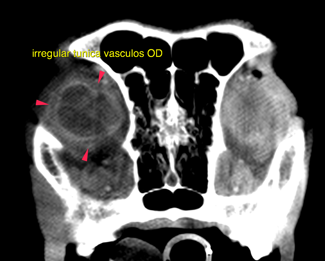
head: Both ocular bulbs appear to be small and deep within the orbita. After contrast administration the vascular tunic of the right ocular bulbus presents an undulating outline. The globoid shape of the left ocular bulb is maintained. However, the vascular tunic presents with double outline on the post contrast series.
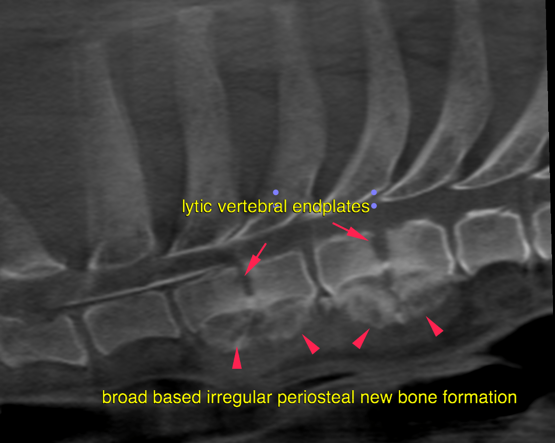
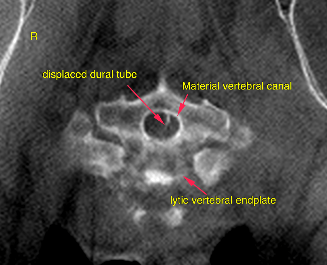
cervical spine – The vertebral endplates of the cervical spine present very mild spondylosis deformans. There is marked periosteal palisading, well delineated, bridging new bone formation at the ventral aspect of the vertebral bodies of T2/3 and T4/5 associated with a moderate circumferential soft-tissue swelling with extension into the ventral epidural space of the vertebral canal. There is moderate moth-eaten osteolysis of the vertebral endplates between T2/3 and T4/5 with peripheral sclerotic margins. The intervertebral disc space is moderately widened. Soft tissue attenuating material is present to the left side of the ventral epidural space with marked mass effect on the spinal cord which is displaced and compressed to the right side level with the intervertebral disc spaces T2/3 and T4/5.