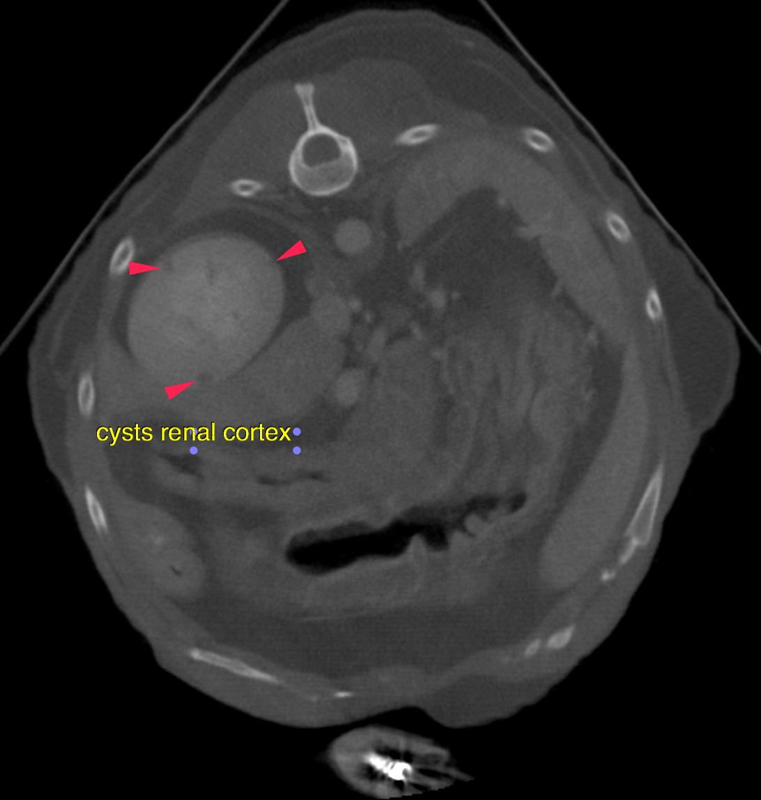
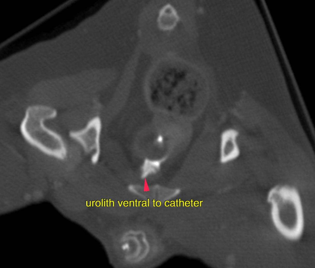
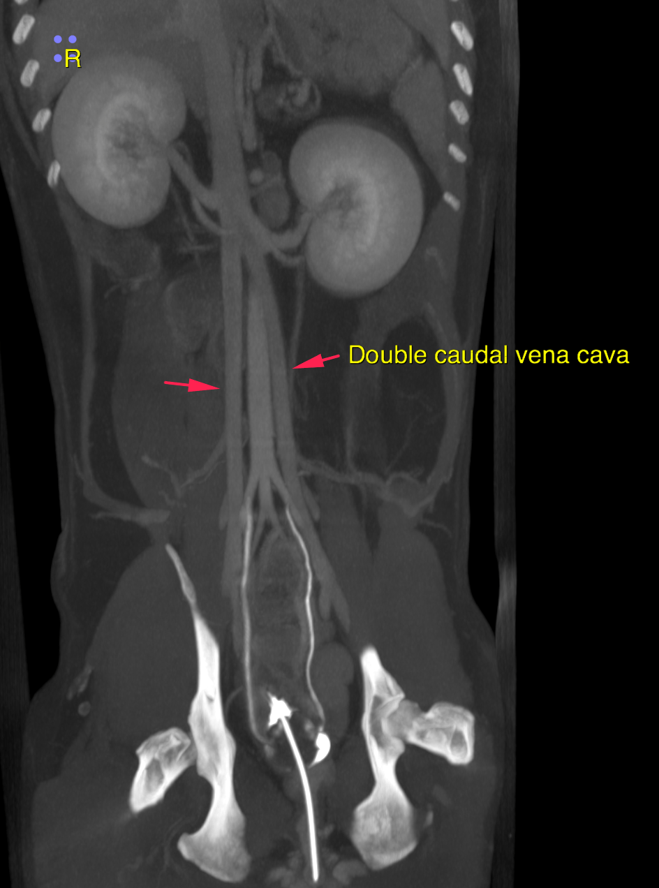
This 10 year old MN Chihuahua has a history of bladder calculi; R/O renal calculi. Crea 0.8 (was 4.5 prior to correction of calculi obstruction)
This 10 year old MN Chihuahua has a history of bladder calculi; R/O renal calculi. Crea 0.8 (was 4.5 prior to correction of calculi obstruction)