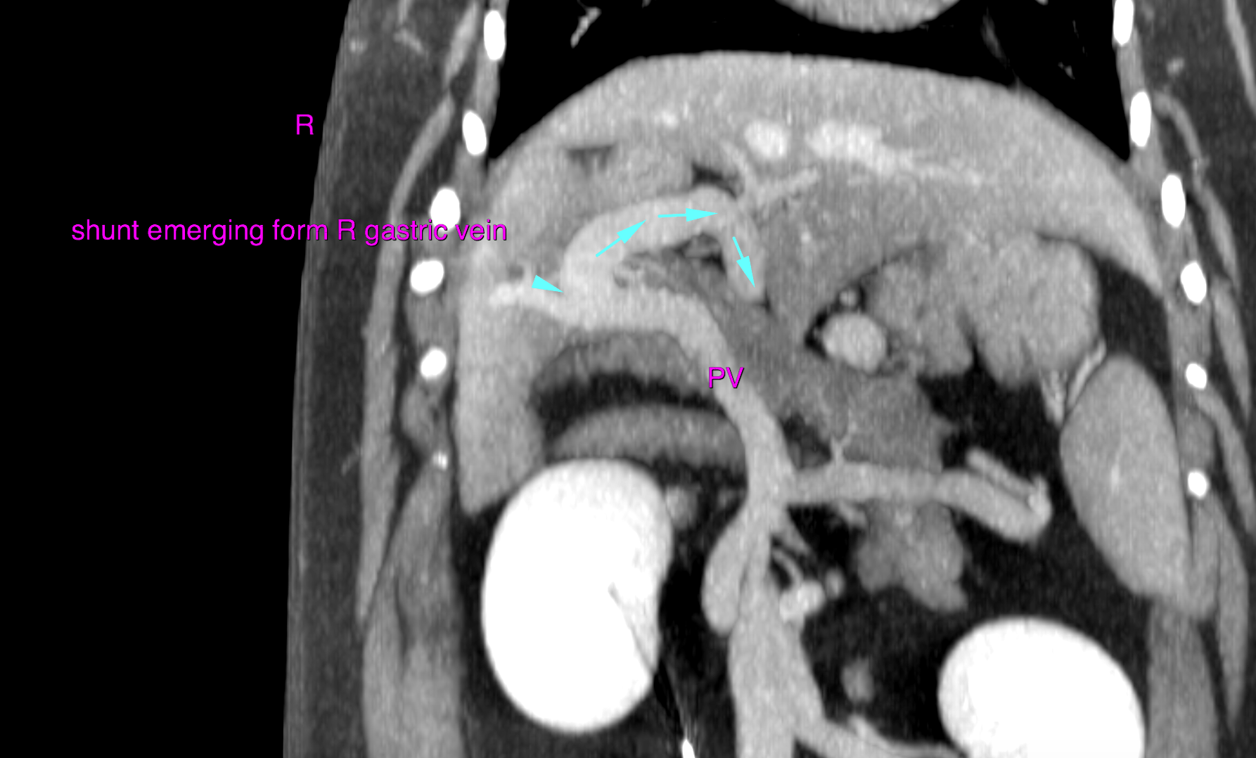
This 1 year old MN Maltese dog presented for bilateral medial luxating patellas. Pre-surgical lab work showed glucoses 121, BUN 6, protein 5.1, albumin 2.3, pre prandial bile acids 336.7
This 1 year old MN Maltese dog presented for bilateral medial luxating patellas. Pre-surgical lab work showed glucoses 121, BUN 6, protein 5.1, albumin 2.3, pre prandial bile acids 336.7