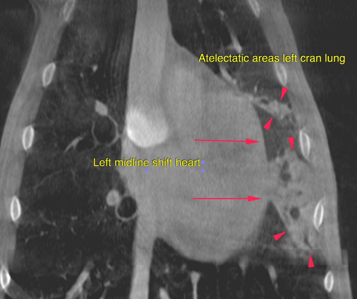
CT – Compression Atelectasis in a 9 year old FS dog

Case Study

CT – Compression Atelectasis in a 9 year old FS dog

This 9 year old FS dog presented with acute coughing

This 9 year old FS dog presented with acute coughing

DX

Incomplete lobar alveolar pattern with volume loss due to compression atelectasis

Image Interpretation

CT of the thorax – 

The caudal compartment of the left cranial lung lobe and the caudal aspect of the cranial compartment adjacent to the heart presents non aerated areas of soft tissue attenuation with air bronchograms, emphasizing the dependent portion of the lobes. Mild volume loss of the left lung with cardiac midline shift to the left side is noted.


Outcome

Less likely tracheitis/laryngitis or treacheal/laryngeal/bronchial collapse. if the clinical signs persist, consider tracheobronchoscopy with bronchoalveolar lavage for further definition.

Patient Information

Patient Name : Jinx Wolf
Gender : Female, Spayed
Species : Canine
Type of Imaging : Ultrasound
Status : Complete
Liz Wuz Here : Yes
Code : 16_00112

Clinical Signs

  • Coughing

Images

bildschirmfoto_2016-08-23_um_12

Clinical Signs

  • Coughing
Skip to content