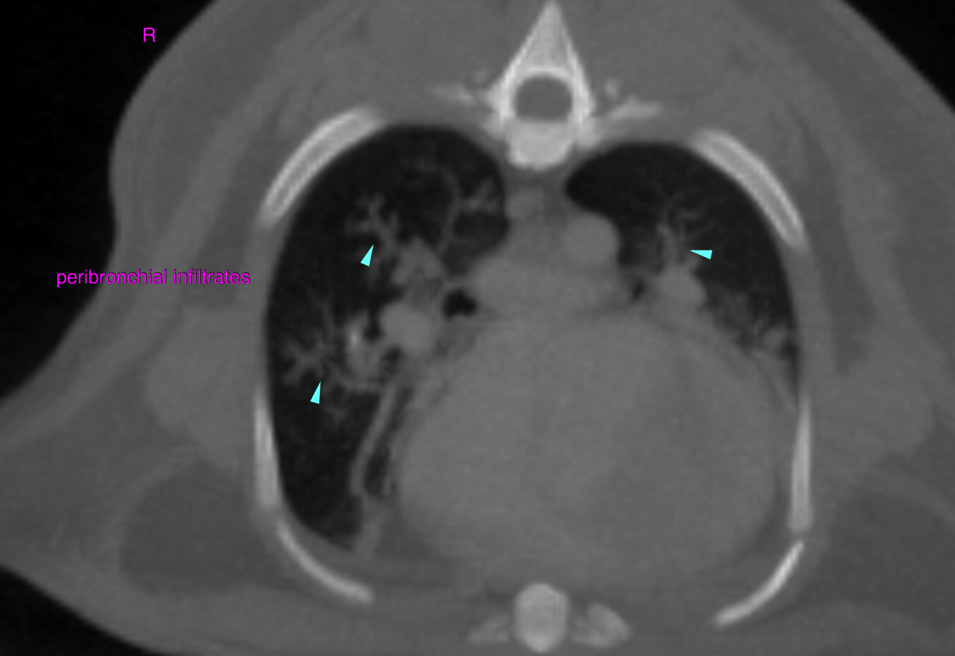
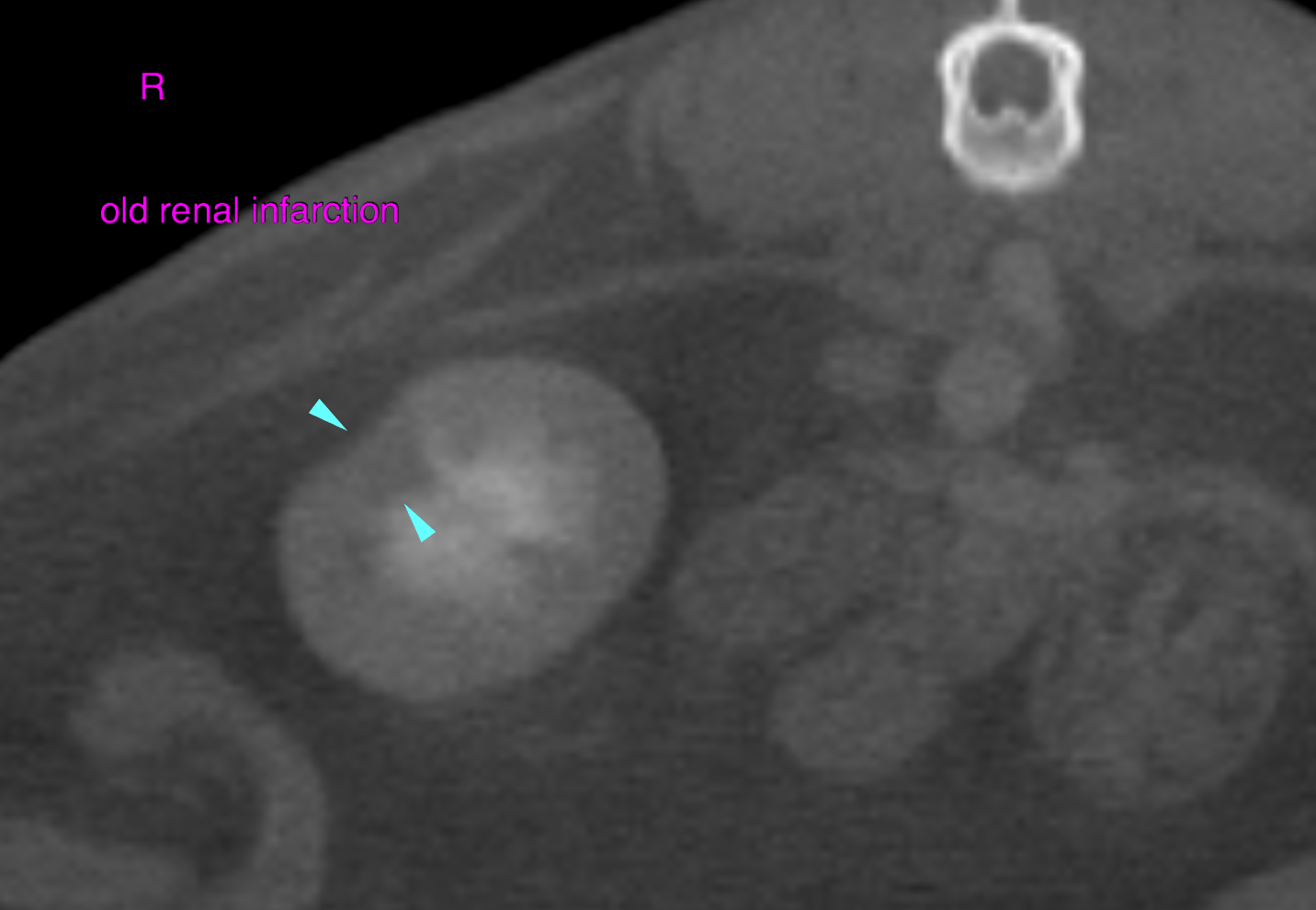
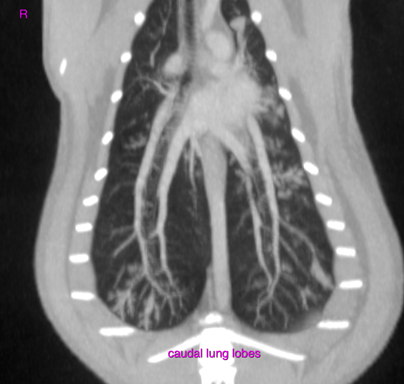
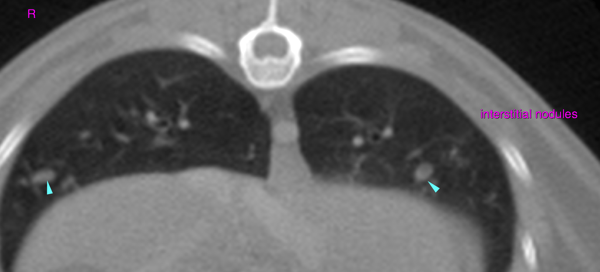
This 14 year old FS DSH cat has a recent history of pulmonary nodule found on a pre-dental workup. New soft tissue opacity noted appearing to arise from the mediastinum. Murmur 3/6, hyperthyroid.
This 14 year old FS DSH cat has a recent history of pulmonary nodule found on a pre-dental workup. New soft tissue opacity noted appearing to arise from the mediastinum. Murmur 3/6, hyperthyroid.