Skip to content
Customize Consent Preferences

We use cookies to help you navigate efficiently and perform certain functions. You will find detailed information about all cookies under each consent category below.

The cookies that are categorized as "Necessary" are stored on your browser as they are essential for enabling the basic functionalities of the site. ... 

Always Active

Necessary cookies are required to enable the basic features of this site, such as providing secure log-in or adjusting your consent preferences. These cookies do not store any personally identifiable data.

No cookies to display.

Functional cookies help perform certain functionalities like sharing the content of the website on social media platforms, collecting feedback, and other third-party features.

No cookies to display.

Analytical cookies are used to understand how visitors interact with the website. These cookies help provide information on metrics such as the number of visitors, bounce rate, traffic source, etc.

No cookies to display.

Performance cookies are used to understand and analyze the key performance indexes of the website which helps in delivering a better user experience for the visitors.

No cookies to display.

Advertisement cookies are used to provide visitors with customized advertisements based on the pages you visited previously and to analyze the effectiveness of the ad campaigns.

No cookies to display.

CT Carcinomatosis with Multi-Centric Spread in a 10 year old MN Siamese cat

Case Study

CT Carcinomatosis with Multi-Centric Spread in a 10 year old MN Siamese cat

This 10 year old MN feline was diagnosed with lymphoma at 2 years of age, was treated with chemotherapy and went into remission. In the last two weeks, a mass was found on right side of jaw. Cytology revealed salivary gland carcinoma.  CBC, chemistry, urinalysis all NSF.

This 10 year old MN feline was diagnosed with lymphoma at 2 years of age, was treated with chemotherapy and went into remission. In the last two weeks, a mass was found on right side of jaw. Cytology revealed salivary gland carcinoma.  CBC, chemistry, urinalysis all NSF.

Image Interpretation

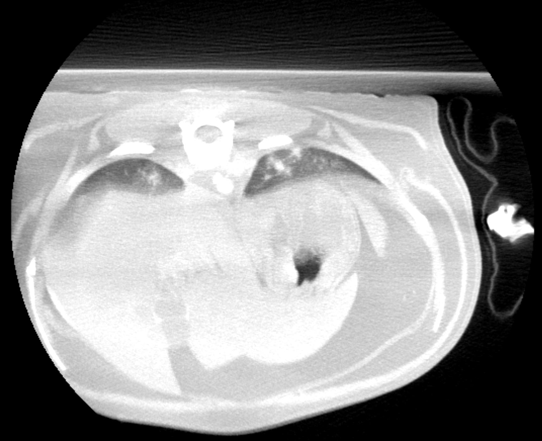
CT of the chest and skull: The chest scan provided covers only the caudal portion of both caudal lung lobes and the cranial abdomen. Within the image aspects of both caudal lung lobes, there are multi focal, soft tissue, attenuating, ill defined interstitial nodules with heterogenous contrast uptake. Atelectatic changes are noted in the dorsal aspects of the lung lobes as the cat was positioned in dorsal recumbency. Within the cranial abdomen, the gallbladder presents bipartite, which is an incidental finding in cats. The pancreas is moderately to severely enlarged and shows multi focal nodular and mass-like lesions expanding the pancreatic capsule. No peripheral fat stranding was noted surrounding the pancreas. The portal lymph nodes both show moderate to severe enlargement, rounding and heterogenous contrast uptake and there are several nodules of varying size within the splenic parenchyma expanding the capsule. Contrast media pooling within the hepatic veins and portal vein, which is likely a function of systemic hypotension under general anesthesia, may also be an indicator of beginning portal hypertension, although no primary changes of the liver were evident from the CT scan. Incidental finding of a mild, emerging spondylosis deformans is noted at multiple sites in the caudal thoracic and cranial lumbar spine. There is a space occupying lesion originating from the mandibular salivary gland on the right side and measuring approximately 2.0 cm in diameter. The lesion shows heterogenous contrast enhancement with predominantly peripheral enhancement and non uniform contrast enhancement in the center of the lesion sparing multiple focal areas. There is moderate enlargement and rounding of the medial retropharyngeal lymph node on the right side with heterogenous contrast uptake. Both tympanic bullae are incompletely filled with soft tissue, attenuating homogenous material. Part of this material on the right side presents with character of a broad based, dorsally attached space-occupying lesion and shows a uniform contrast enhancement. Both tympanic bulla walls show moderate, uniform thickening. There is mild swelling of the nasopharyngeal soft tissues, medial to both tympanic bullae. Bilateral chronic otitis media with poly formation on the presumed right side.

DX

Findings are compatible with a malignant neoplasia with multi centric spread.

Outcome

From the CT scan, it is not possible to determine the primary lesion of either the mandibular salivary gland adenocarcinoma or the pancreas. As the pancreatic findings are compatible with an adenocarcinoma with metastatic spread and this could be the primary lesion in this case. The overall prognosis is poor as a carcinomatosis with multi centric metastatic spread is evident from the CT scans.
**Left and right was shifted due to erroneous set up at least for the chest and cranial abdomen.

Patient Information

Patient Name : Nutley - Alpine Veterinary Hospital
Gender : Male, Neutered
Species : Feline
Type of Imaging : Ultrasound
Status : Complete

History

  • Neoplasia

Exam Finding

  • Masses

Images

skull_postskull_post_arrowedskull_post2_arrowedthorax_far_postthorax_far_post2_arrowedthorax_far_post2thorax_far_post3_arrowed