CT of the elbows and shoulders –
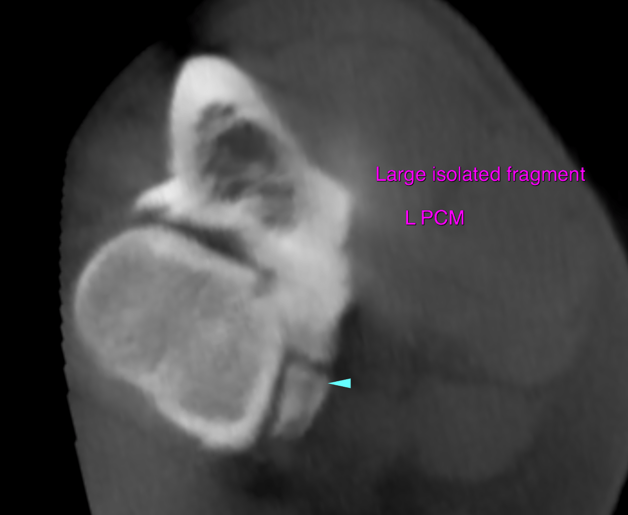
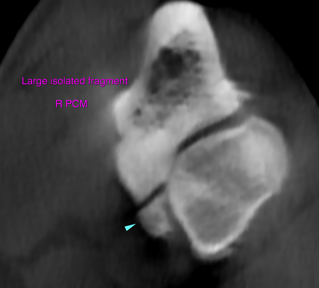
Elbows: Both medial coronoid processes present a large isolated bone fragment of approximately 6 mm side length. The base of the medial coronoid processes reveals increased bone sclerosis. Moderate narrowing and incongruity of the radioulnar incisure is noted. A mild radioulnar step formation with a long ulna is seen bilaterally. The humeroulnar joint spaces are mildly asymmetric. The subchondral bone of the medial humeral condyle presents focal sclerosis opposed to the fragments resepctively. A moderate amount of osteophytes is seen at the periarticular margins of the left and right elbow joint.
Shoulders: Both shoulder joints present within normal limits