CT – Bilateral biceps tendovaginitis in a Labrador Retriever with shifting leg lameness

Case Study

CT – Bilateral biceps tendovaginitis in a Labrador Retriever with shifting leg lameness

This Labrador Retriever dog presented with shifting leg lameness that was non-responsive to chiropractic care or acupuncture. Normal MRI.

This Labrador Retriever dog presented with shifting leg lameness that was non-responsive to chiropractic care or acupuncture. Normal MRI.

DX

Indirect signs of chronic bicipital tendovaginitis with shoulder joint osteoarthritis: moderate on the left, mild on the right side

Image Interpretation

CT of the cervical spine, shoulders and elbows – 

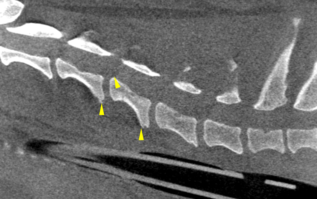
Cervical spine: Mild osteophyte formations emerging from the neighboring vertebral endplates of C4/C5 and C5/C6 consistent with emerging Spondylosis deformans are noted along with mild craniodorsal tipping of the vertebral body of C5 and mild intervertebral disc protrusion.

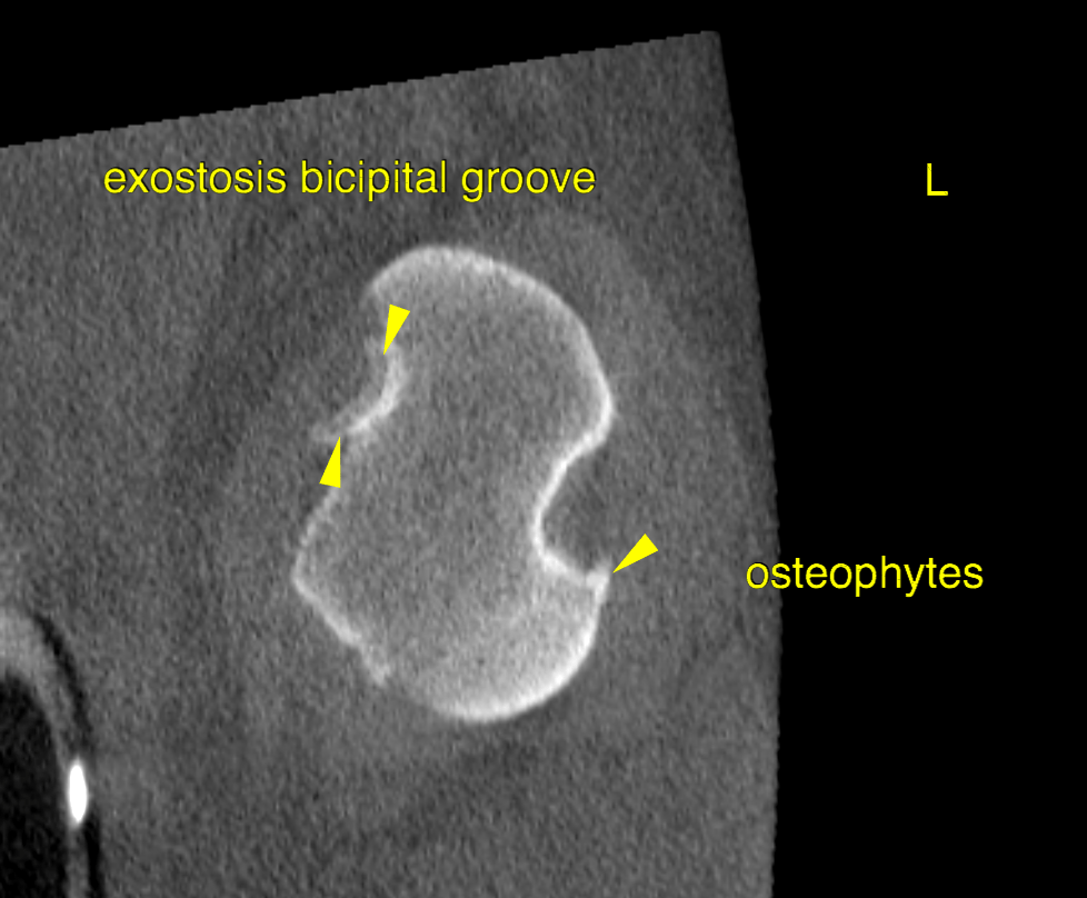
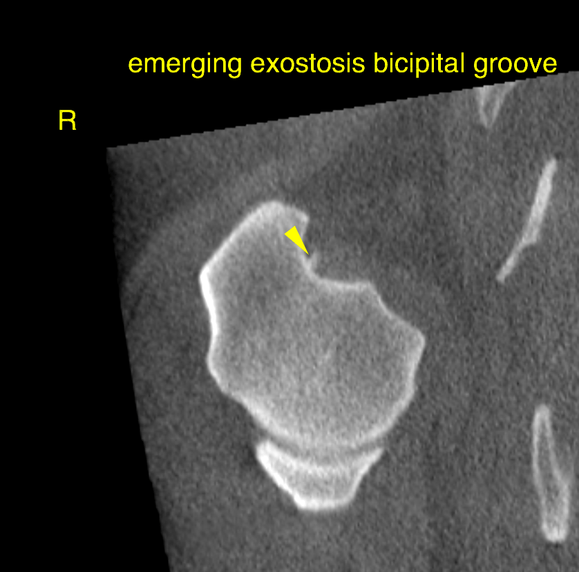
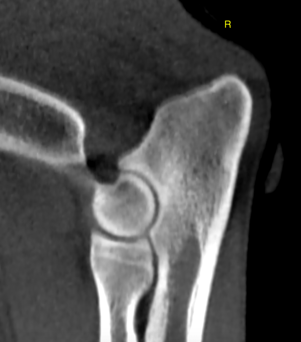
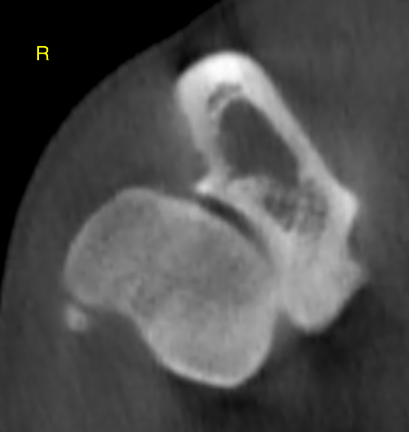
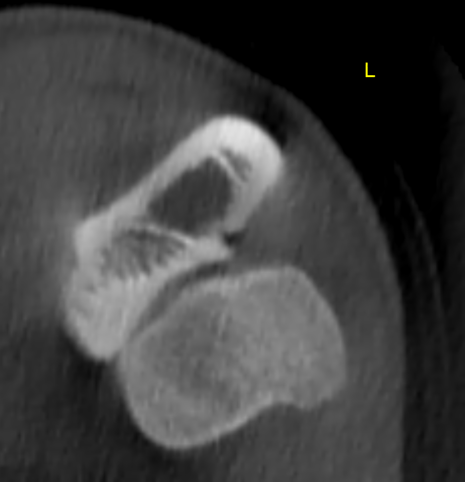
Shoulders: The left shoulder reveals moderate degenerative joint disease with osteophytosis and subchondral bone sclerosis and joint effusion. Moreover there are indirect signs of chronic biceps tendovaginitis with marked semicircular bony exostosis and sclerosis within the intertubercular groove of the biceps tendon.

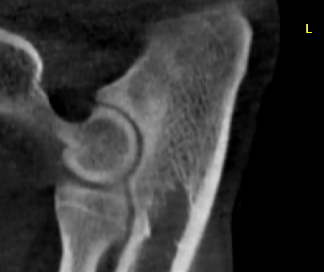
Elbows: The medial coronoid process of both elbows reveals increased sclerosis with loss of the regular trabecular bone pattern. The delineation of the medial coronoid processes is smooth respectively. There is no fragment or fissure line identified. The joint surfaces are congruent. Emerging oesteophytes are noted at the periarticular margins.

Outcome

No evidence of relevant elbow dysplasia. No evidence of compressive myelopathy throughout the cervical spine. The computed tomographic findings are suggestive for bilateral biceps tendovaginitis. which matches a chronic/intermittent shifting lameness that is mild to moderate in
degree and dependent on the work load. Medium to large breed dogs older than 5-6
years are typically affected whereas clinical signs of elbow dysplasia usually peak
around an age of 6 to 24 months.

Patient Information

Patient Name : Archie Adler
Species : Canine
Type of Imaging : Ultrasound
Status : Complete
Liz Wuz Here : Yes
Code : 16_00051

Clinical Signs

  • Lameness

Images

bildschirmfoto_2016-03-08_um_20bildschirmfoto_2016-03-08_um_20bildschirmfoto_2016-03-09_um_09bildschirmfoto_2016-03-09_um_09bildschirmfoto_2016-03-09_um_09bildschirmfoto_2016-03-09_um_09bildschirmfoto_2016-03-09_um_09

Clinical Signs

  • Lameness
Skip to content