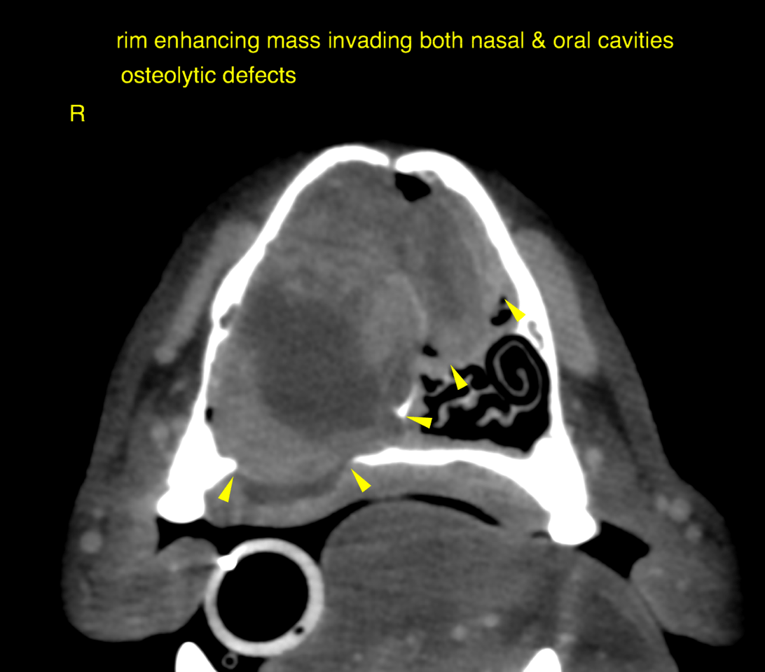
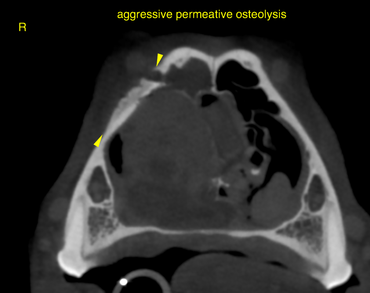
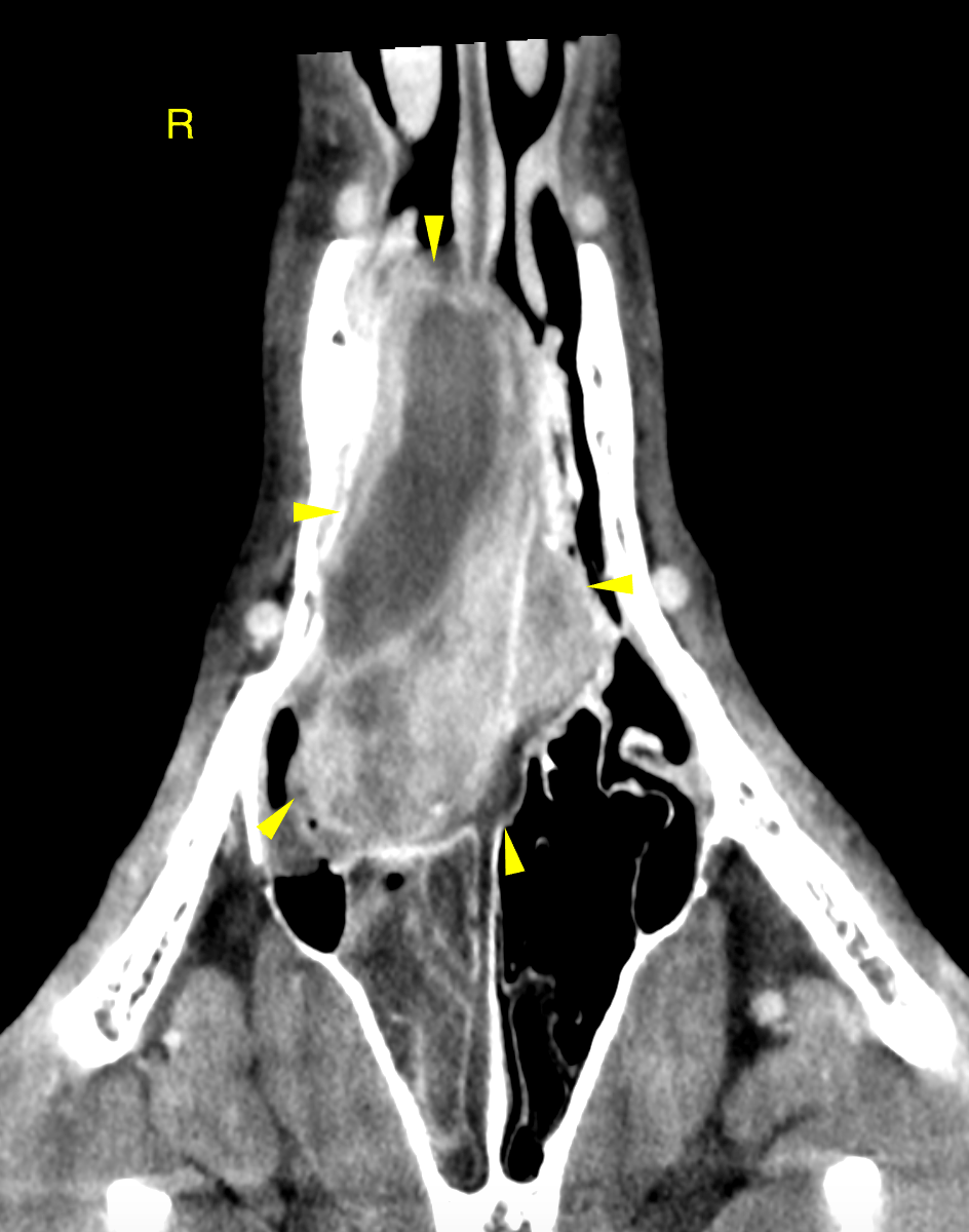
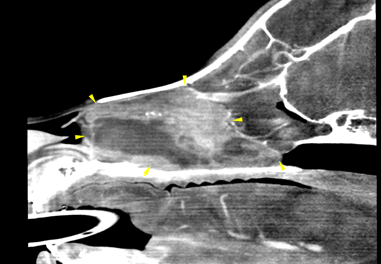
This 7 year old MN Australian Shepherd has a 2 week history of epistaxis when playing, nose seems congested.
Physical exam: unable to pass air in nasal passages, open mouth breathing
CBC/Chem: ALB, ALT, ALKP high; Glob, amylase low
This 7 year old MN Australian Shepherd has a 2 week history of epistaxis when playing, nose seems congested.
Physical exam: unable to pass air in nasal passages, open mouth breathing
CBC/Chem: ALB, ALT, ALKP high; Glob, amylase low