CT – Advanced degenerative joint disease (DJD) with medial coronoid pathology and kissing lesion in a 6 year old MN Bernese Mountain Dog

Case Study

CT – Advanced degenerative joint disease (DJD) with medial coronoid pathology and kissing lesion in a 6 year old MN Bernese Mountain Dog

This 6 year old MN Bernese Mountain Dog presented with right front lameness. Painful on palpation, particularly the medial distal humerus and proximal tibia. R/O fracture vs neoplasia.

This 6 year old MN Bernese Mountain Dog presented with right front lameness. Painful on palpation, particularly the medial distal humerus and proximal tibia. R/O fracture vs neoplasia.

DX

• Advanced degenerative joint disease • Medial coronoid pathology with fragmentation of the medial coronoid process, kissing lesion(s) within the medial humeral condyle and complex elbow joint incongruity • Possible chronic flexor enthesiopathy • Mineralization in the joint capsule right elbow joint

Image Interpretation

CT of the right elbow – 

A fissure line at the tip of the medial coronoid process separates a demineralized bone fragment of 3.5 mm diameter. The base of the medial coronoid process shows a moderately heterogeneously increased sclerosis. Moderate regional sclerosis of the subchondral bone of the medial humeral condyle is noted on the opposite side of the isolated coronoid fragment and associated with a small subchondral bone defect of approximately 2 mm diameter and 1 mm depth. The subchondral bone of the medial humeral condyle and trochlea notch of the ulna is moderately irregular with small crescent shaped indentations. A mild radioulnar step formation with a long ulna is noted as well as crescent shaped asymmetry of the humeroulnar joint space. In the area of the insertion of the joint capsule at the cranial the humerus there is well defined round, 3.5mm in diameter mineral attenuating structure visible.


Outcome

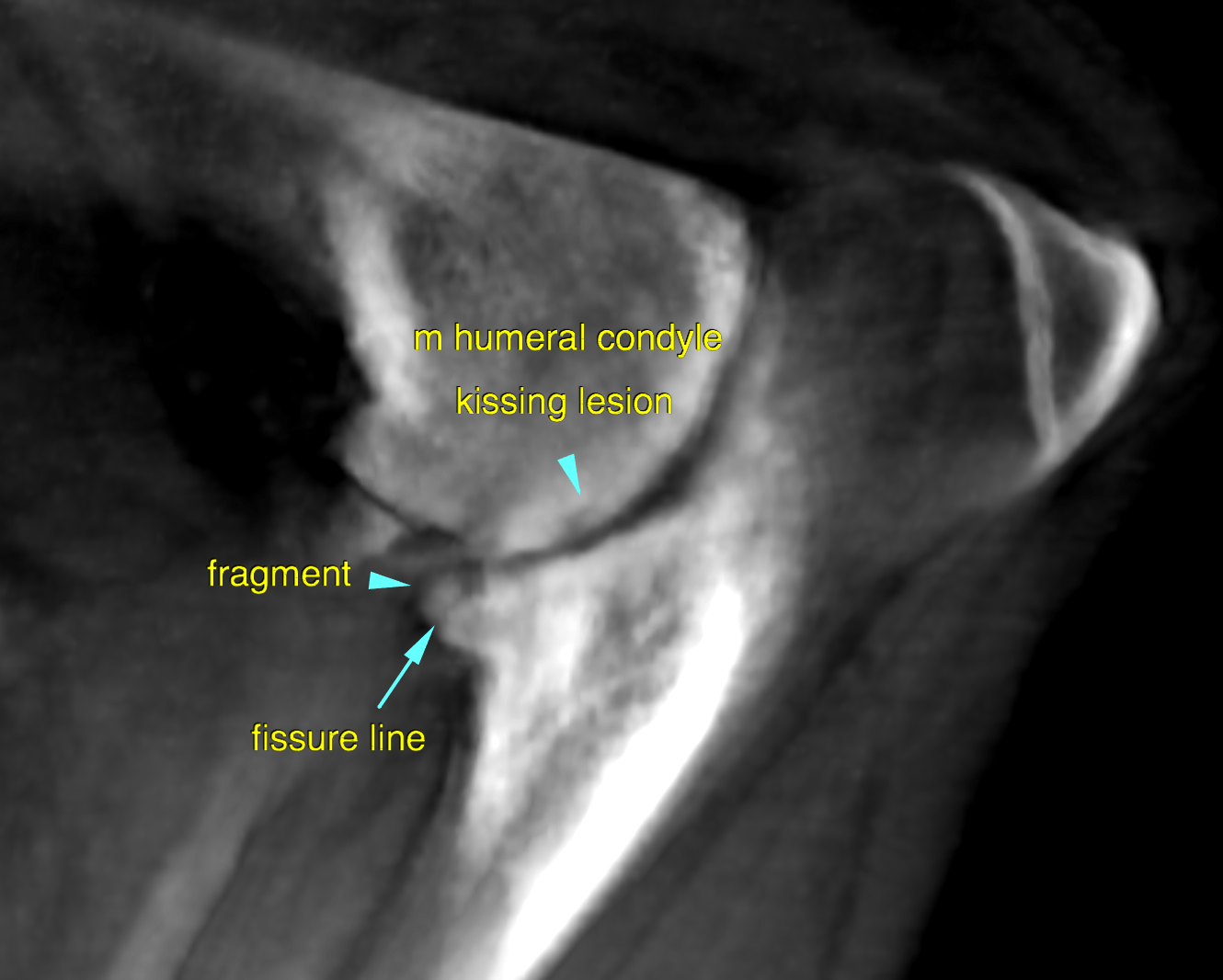
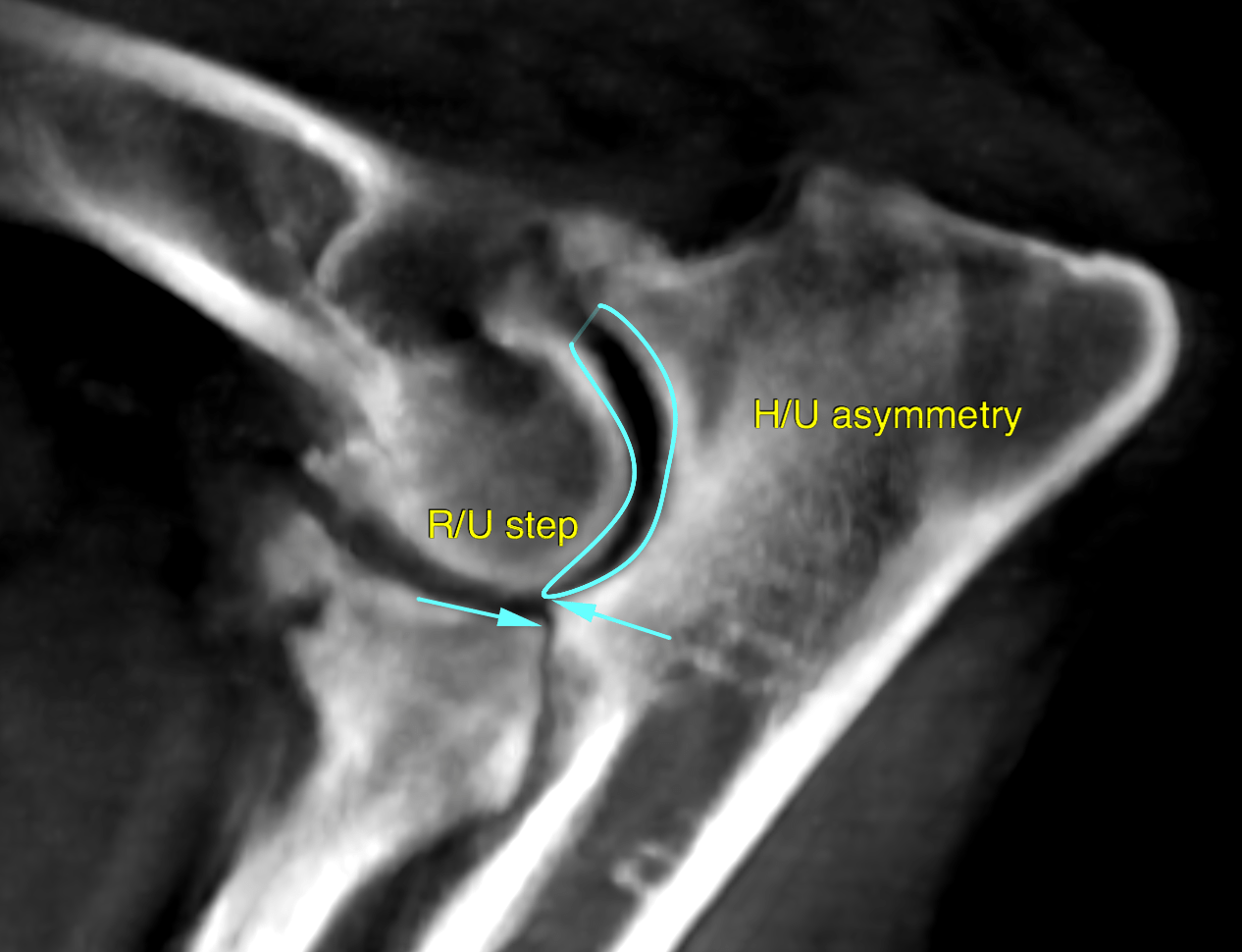
The findings are compatible with chronic DJD of the right elbow due to a fragmented medial coronoid process. The osteoarthrosis and the findings in the medial humeral condyle are chronic secondary changes to the primary coronoid disease. Advanced cartilage breakdown – especially within the medial joint compartment is assumed. Still arthroscopy is recommended to revise the joint and remove the fragment of the medial coronoid process and prevent further damage.

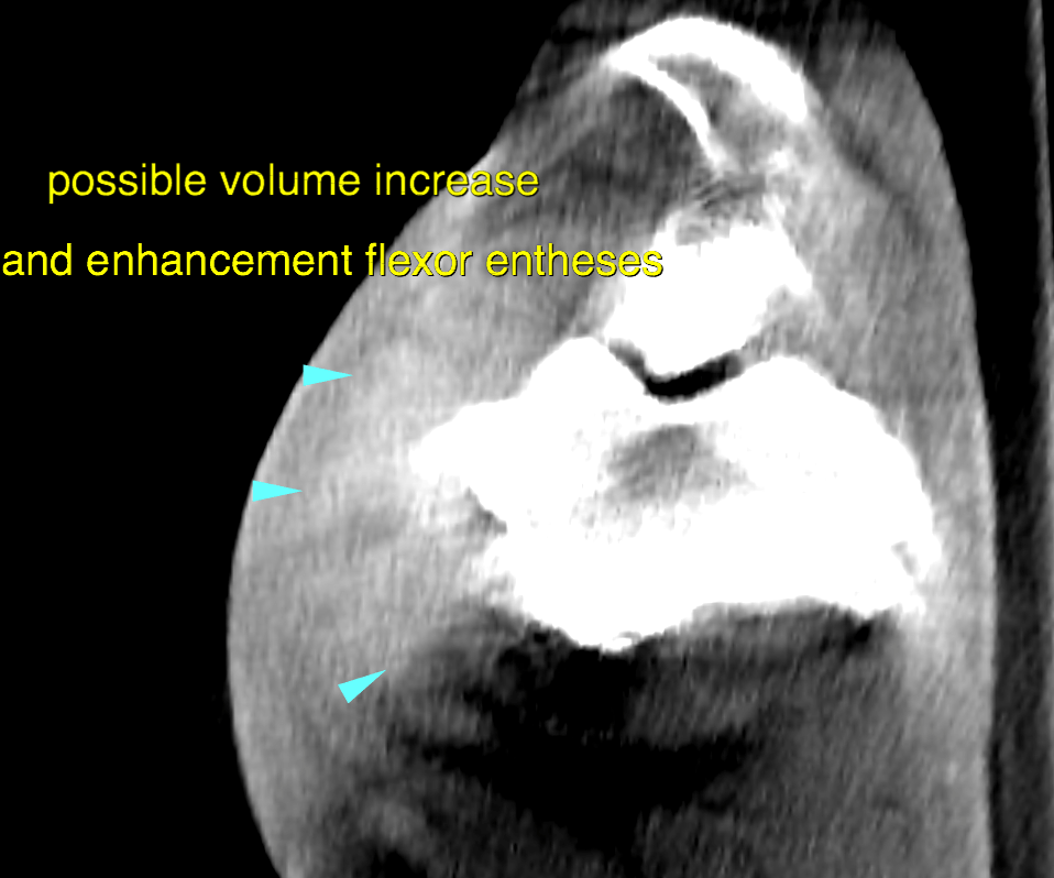
The overall prognosis for the joint is guarded and a lifelong treatment with NSAID’s or other palliative pain management will likely be necessary. Complementary physical therapy to address the possible flexor enthesiopathy.

The mineralization in the joint capsule is compatible with synovial osteochondromatosis and an incidental finding.

Patient Information

Patient Name : Matisse Lacy/Neel
Gender : Male, Neutered
Species : Canine
Type of Imaging : Ultrasound
Status : Complete
Liz Wuz Here : Yes
Code : 16_00101

Clinical Signs

  • Lameness

Exam Finding

  • Pain

Images

bildschirmfoto_2016-08-16_um_11bildschirmfoto_2016-08-16_um_11bildschirmfoto_2016-08-16_um_11

Clinical Signs

  • Lameness
Skip to content