CT – Acute Lumbosacral Disc Extrusion in a 9 year old Old English Sheepdog

Case Study

CT – Acute Lumbosacral Disc Extrusion in a 9 year old Old English Sheepdog

The patient presented with acute grade 3 paraparesis with neuroanatomic localization at L4-S3 and severe pain. 

The patient presented with acute grade 3 paraparesis with neuroanatomic localization at L4-S3 and severe pain. 

DX

Lumbosacral nucleus pulpous extrusion leading to severe lumbosacral stenosis with compressive neuropathy of the Cauda equina.

Image Interpretation

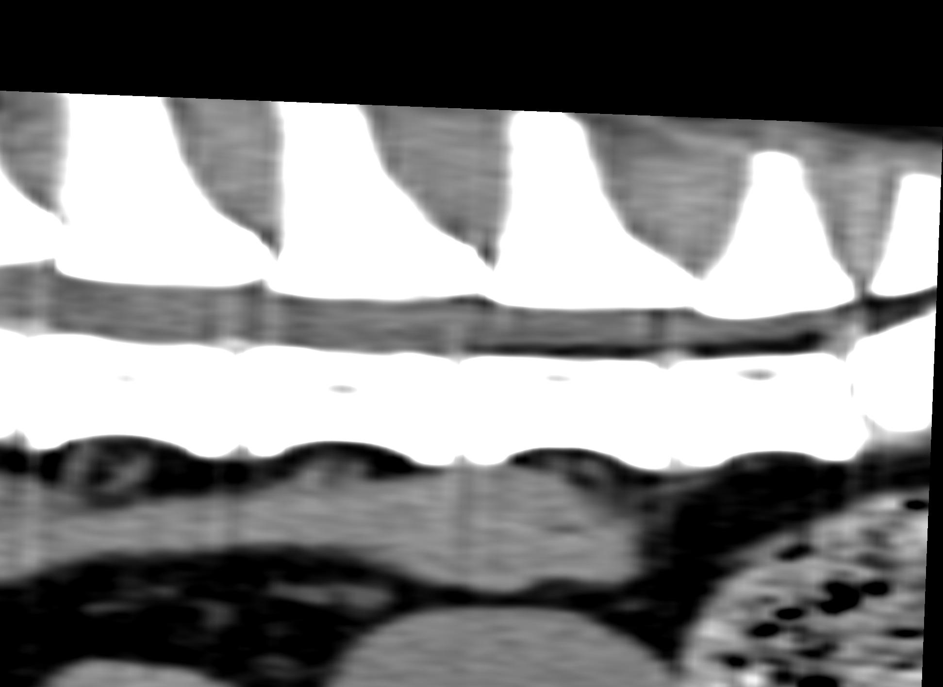
A large volume of hyperattenuating disc material was seen ventrally within the epidural space of the lumbosacral vertebral canal. The material presented cranial and caudal expansion. 90% of the vertebral canal diameter was occupied by the herniated material. There was significant dorsal displacement and compression of the cauda equina nerve roots. The lumbosacral neuroforamina presented no obstruction. Mild noncompresive disc protrusions were seen along the lumbar spine.

Outcome

The dog underwent dorsal laminectomy where the nucleus pulpous extrusion was confirmed and revised.

Patient Information

Gender : Female, Spayed
Species : Canine
Type of Imaging : Ultrasound

Clinical Signs

  • Hind limb paralysis
  • Neurologic signs
  • Weakness

History

  • Weakness

Exam Finding

  • Neurologic signs
  • Weakness

Images

ws_nativ_3ws_nativ_3ws_nativ_3ws_nativ_3

Clinical Signs

  • Hind limb paralysis
  • Neurologic signs
  • Weakness
Skip to content