Common bile duct (CBD) and duodenal papilla obstructive calculi and gallbladder calculi in a 12 year old MN DSH cat

Case Study

Common bile duct (CBD) and duodenal papilla obstructive calculi and gallbladder calculi in a 12 year old MN DSH cat

A 12-year-old NM DSH cat was presented for evaluation of inappetence for 3 days.  Physical examination showed severe dental disease, gallop arrhythmia, and dull mentation. Abnormalities on serum biochemistry were hyperglycemia (270), elevated BUN (39) and ALT (473) activity, and severely elevated total protein (9.6) and globulins (6.8). Radiographs revealed hepatomegaly and a gas filled colon and small intestine.
A 12-year-old NM DSH cat was presented for evaluation of inappetence for 3 days.  Physical examination showed severe dental disease, gallop arrhythmia, and dull mentation. Abnormalities on serum biochemistry were hyperglycemia (270), elevated BUN (39) and ALT (473) activity, and severely elevated total protein (9.6) and globulins (6.8). Radiographs revealed hepatomegaly and a gas filled colon and small intestine.
DX

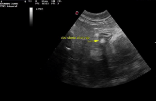
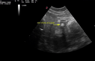
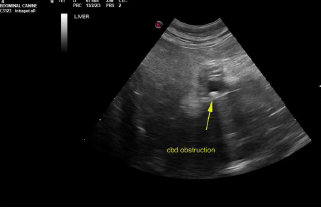
Common bile duct and duodenal papilla obstructive calculi. Gallbladder calculi.

Sonographic Differential Diagnosis

Common bile duct and duodenal papilla obstructive calculi.
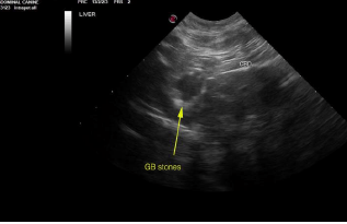
Gallbladder calculi.
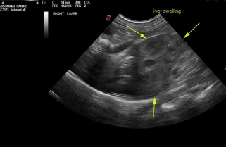
Mild heterogenous hepatic changes. Cholangiohepatitis pattern with lobar swelling at the caudate process.
Age related renal changes with cortical cysts.
Urinary bladder with debris.

Image Interpretation

Liver in this patient presented heterogenous, irregular caudate process with capsular expansion. Recommend ultrasound guided FNA of this portion of the liver. Hepatoma possible, yet relatively rare in cats. Gallbladder presented thickened echogenic wall with calculi. Common bile duct was dilated with focal calculi. Near the duodenal papilla another was imbedded within the duodenal papilla. 0.5 cm calculus noted at the duodenal papilla causing posthepatic obstruction.

Outcome

Surgical intervention with removal of the common bile duct and gallbladder calculi. Removal of the gallbladder likely ideal in this patient given the echogenic wall which usually indicates fibrosis. Sampling of the caudate process of the liver could also be considered first as a screening measure or direct sampling at surgery. This portion of the liver could also be removed directly; appears to be resectable.

Clinical Differential Diagnosis

Liver – cholangio-hepatitis complex, neoplasia, granulomatous disease
Gall bladder – cholecystitis, obstruction (lith, neoplasia, duodenal/pancreatic disease
Pancreas – pancreatitis, neoplasia
Systemic – FIP, hyperthyroidism, multiple myeloma

Patient Information

Patient Name : Clark Rosswog/Easter AH
Gender : Male, Neutered
Species : Feline
Type of Imaging : Ultrasound
Status : Complete
Liz Wuz Here : Yes

Clinical Signs

  • Anorexia

Exam Finding

  • Arrhythmia
  • Dental disease
  • Hepatomegaly
  • Mentally dull

Images

screen_shot_2016-04-30_at_12screen_shot_2016-04-30_at_12screen_shot_2016-04-30_at_12screen_shot_2016-04-30_at_12screen_shot_2016-04-30_at_12

Blood Chemistry

  • ALT (SGPT), High
  • BUN high
  • Globulin, High
  • Glucose, High
  • Total Protein, High

Clinical Signs

  • Anorexia
Skip to content