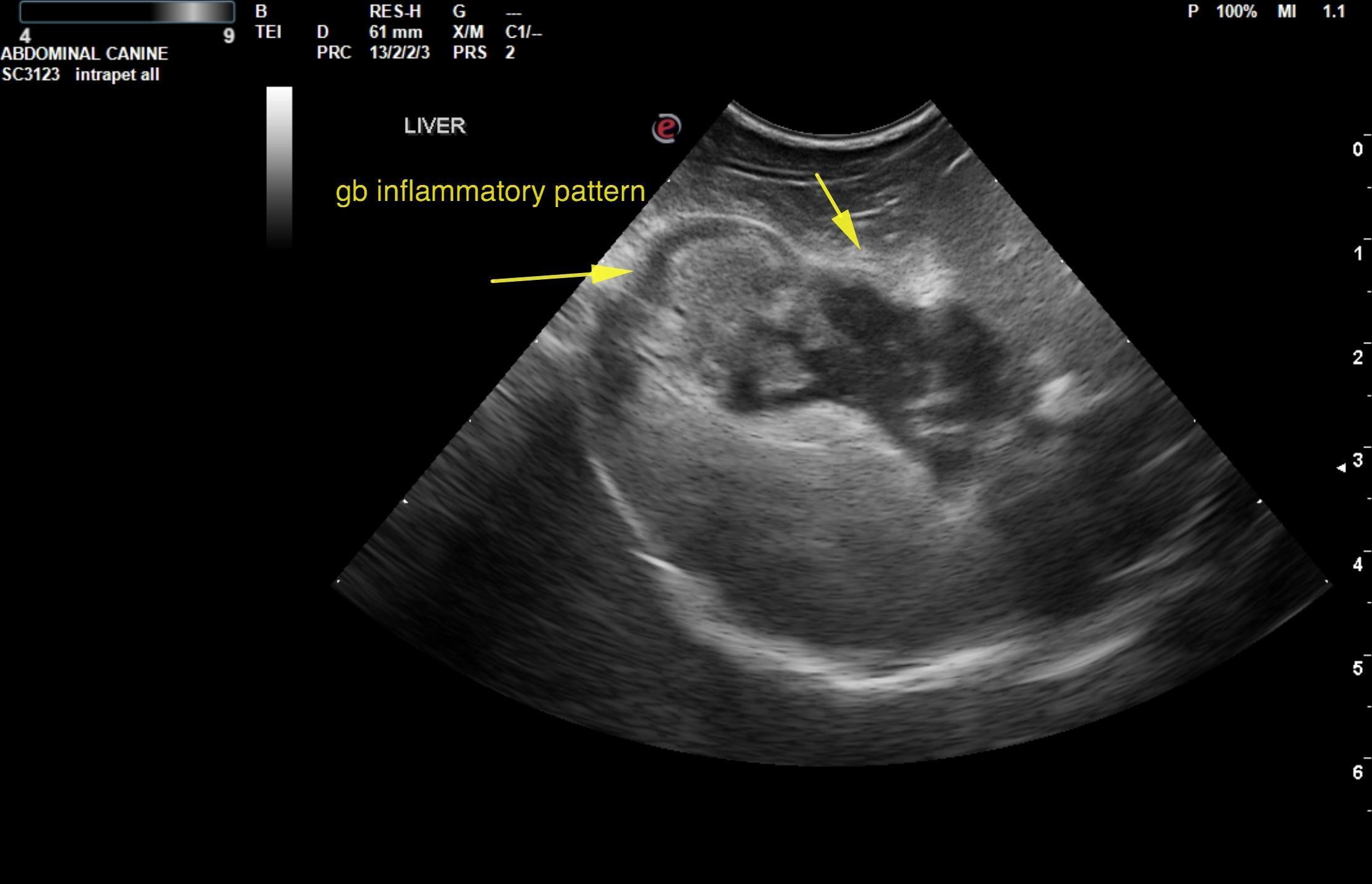
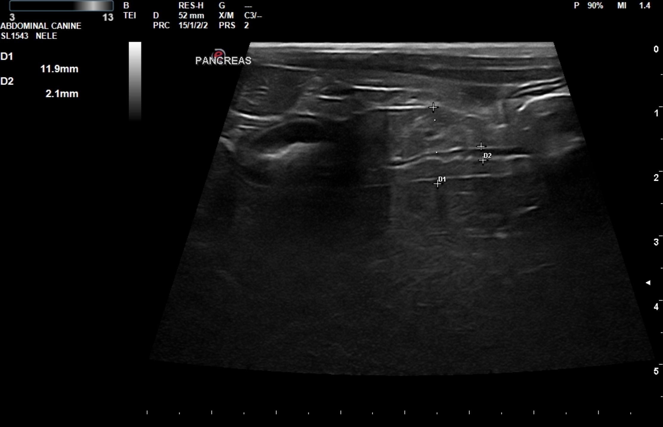
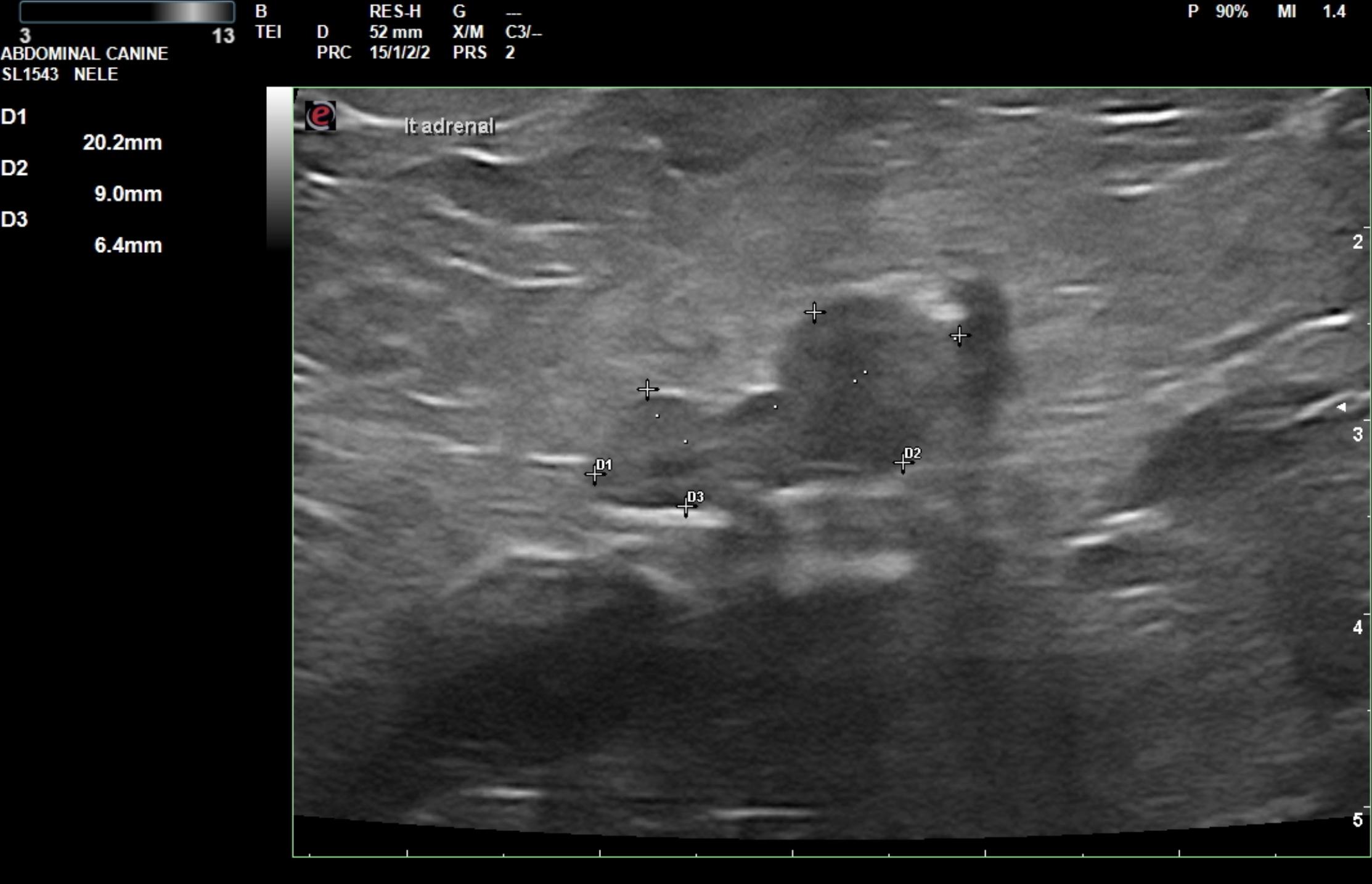
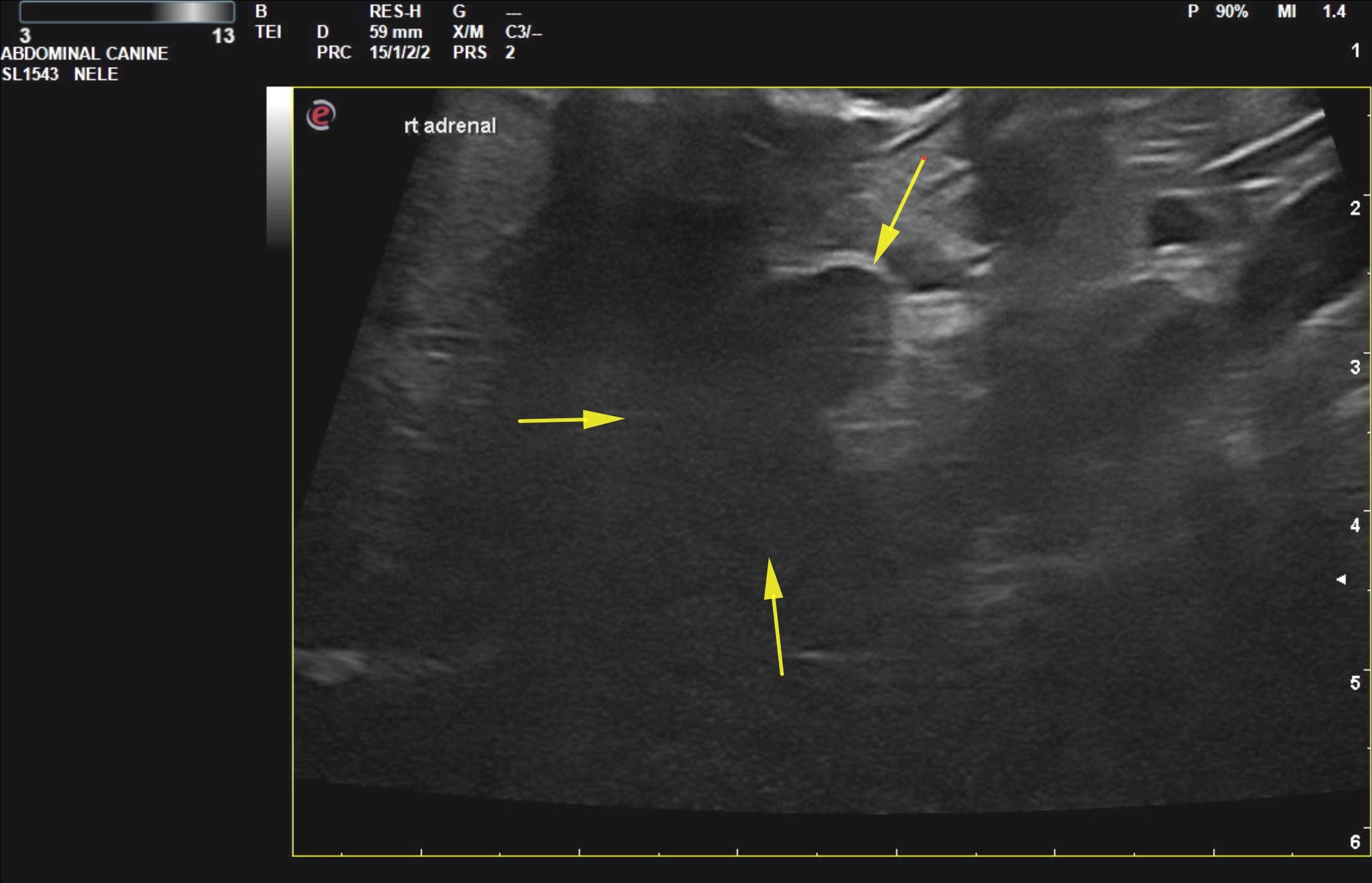
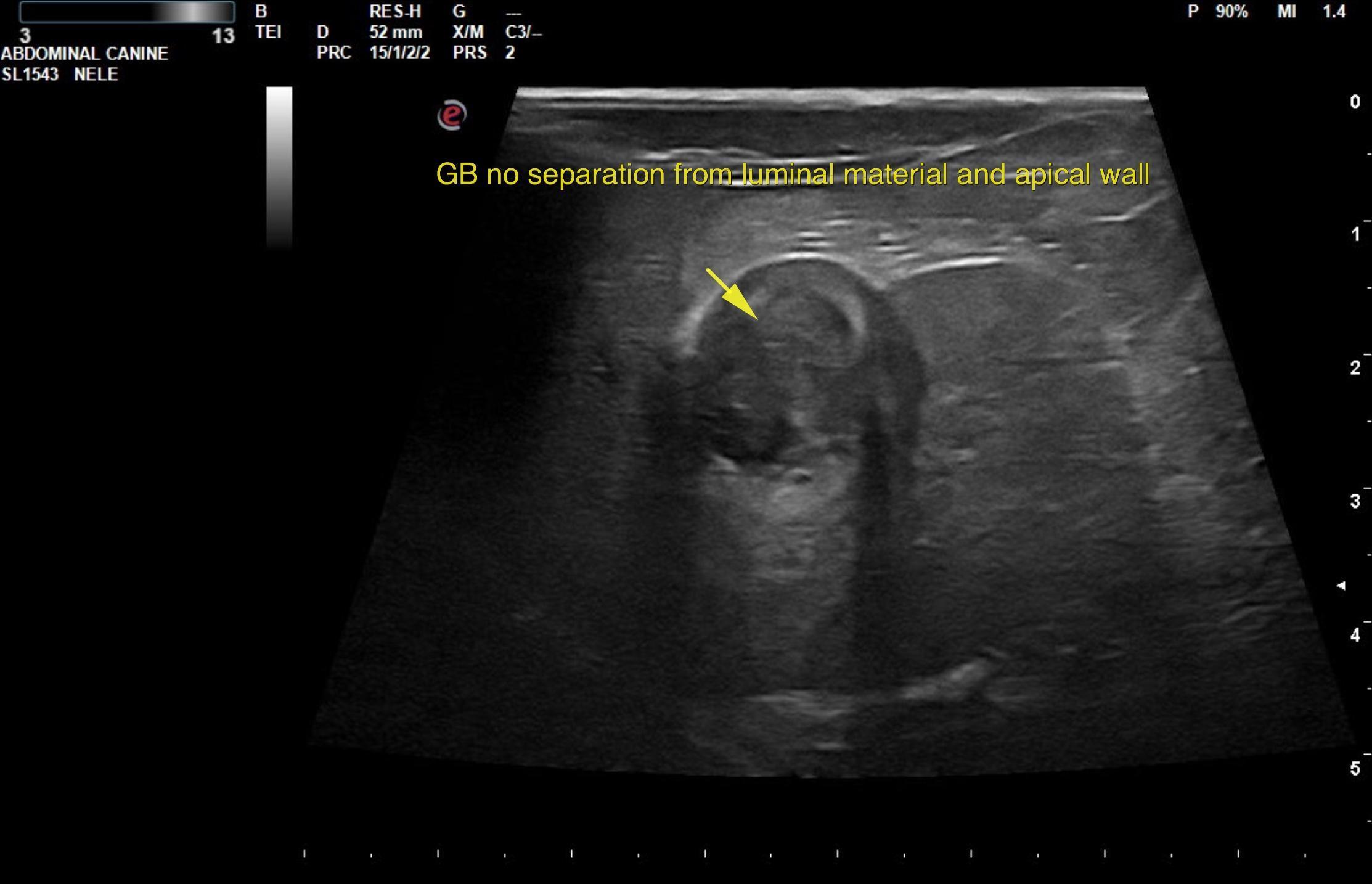
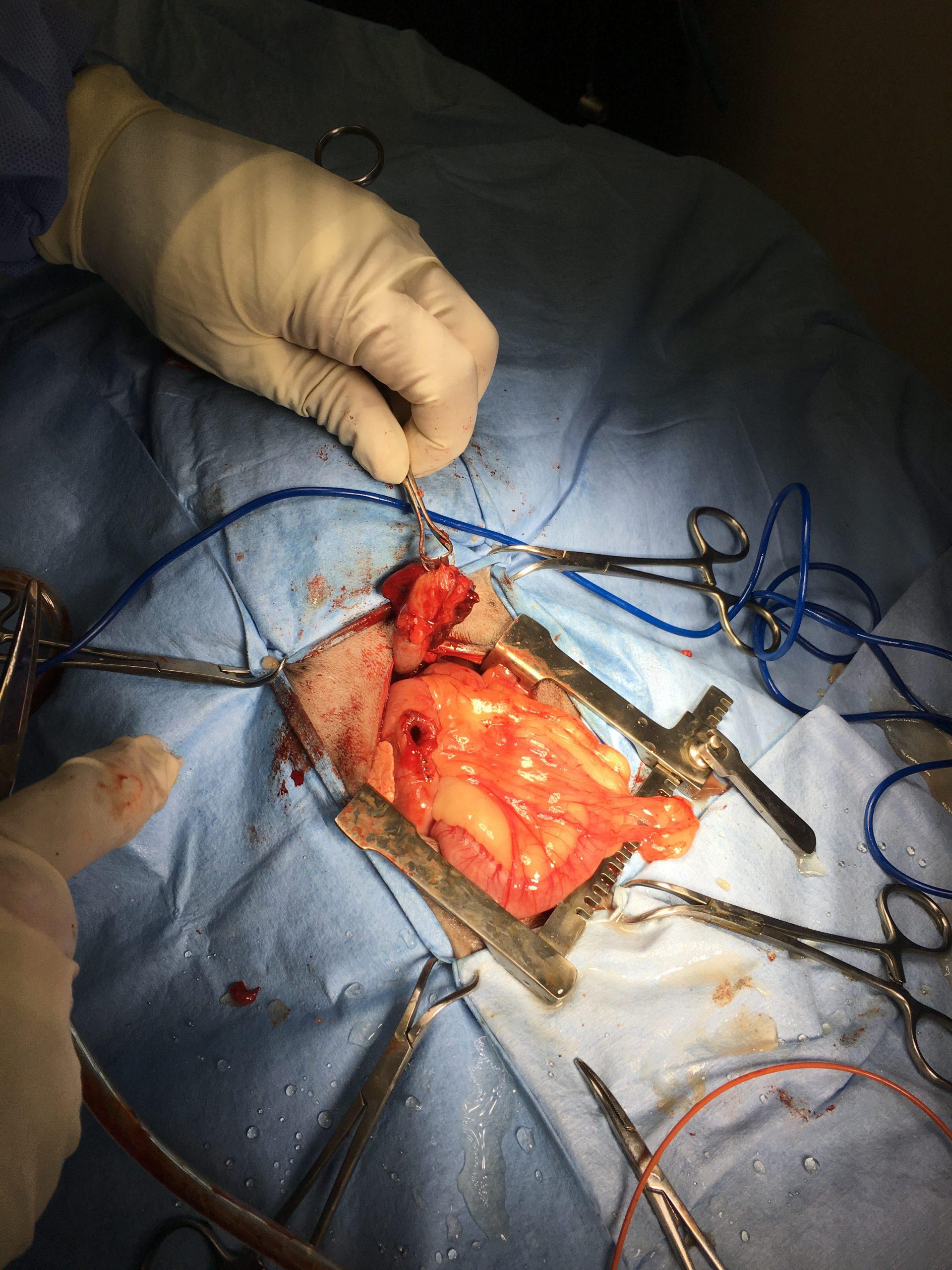
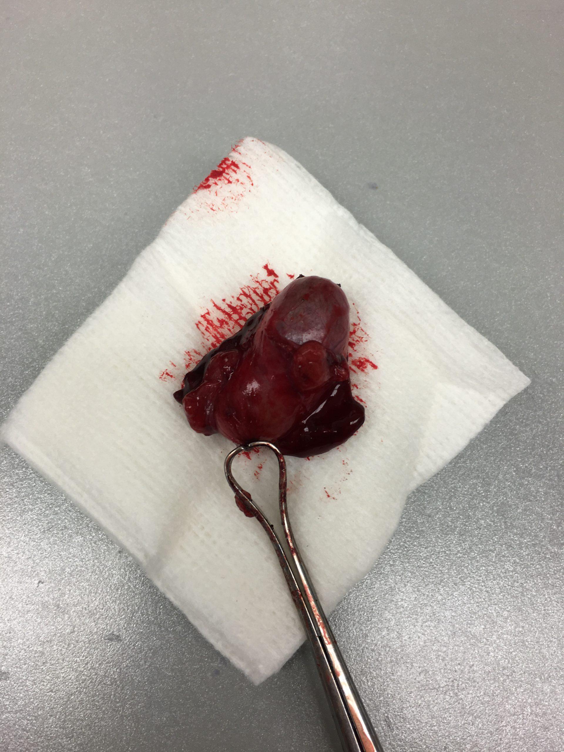
A 10-year-old MN Chihuahua with a history of diabetes and severe pancreatitis was presented for evaluation of PU/PD. Urinalysis showed glycosuria and proteinuria with a normal specific gravity (1.039). Abnormalities on serum biochemistry were elevated cholesterol (1257), triglycerides (2485), PSL (229), ALT (360), ALP (5307), GGT (28), BUN (36), and glucose (275).
A 10-year-old MN Chihuahua with a history of diabetes and severe pancreatitis was presented for evaluation of PU/PD. Urinalysis showed glycosuria and proteinuria with a normal specific gravity (1.039). Abnormalities on serum biochemistry were elevated cholesterol (1257), triglycerides (2485), PSL (229), ALT (360), ALP (5307), GGT (28), BUN (36), and glucose (275).