A 7-year-old FS Labrador Retriever dog with recent history of resolved corneal laceration and Leptosporosis vaccination was presented for intermittent vomiting and diarrhea. The only significant abnormality on physical examination was mild icterus. PCV/TP was 36/5.8 and 4DX negative. The patient was treated with I.V, fluids, antibiotics, anti-emetics, gastroprotectants, and vitamin K. 24 hours later the patient was nauseous and icteric.
A 7-year-old FS Labrador Retriever dog with recent history of resolved corneal laceration and Leptosporosis vaccination was presented for intermittent vomiting and diarrhea. The only significant abnormality on physical examination was mild icterus. PCV/TP was 36/5.8 and 4DX negative. The patient was treated with I.V, fluids, antibiotics, anti-emetics, gastroprotectants, and vitamin K. 24 hours later the patient was nauseous and icteric.
Case Study
Chronic active hepatitis in a 7 year old FS Labrador Retriever dog
Sonographic Differential Diagnosis
Chronic active hepatitis, cholangiohepatitis presentation with portal hypertension. Suspect infectious disease such as Leptospirosis or hepatotoxin. Ascites likely owing to portal hypertension. Chronic interstitial renal pattern. Ascites.
Image Interpretation
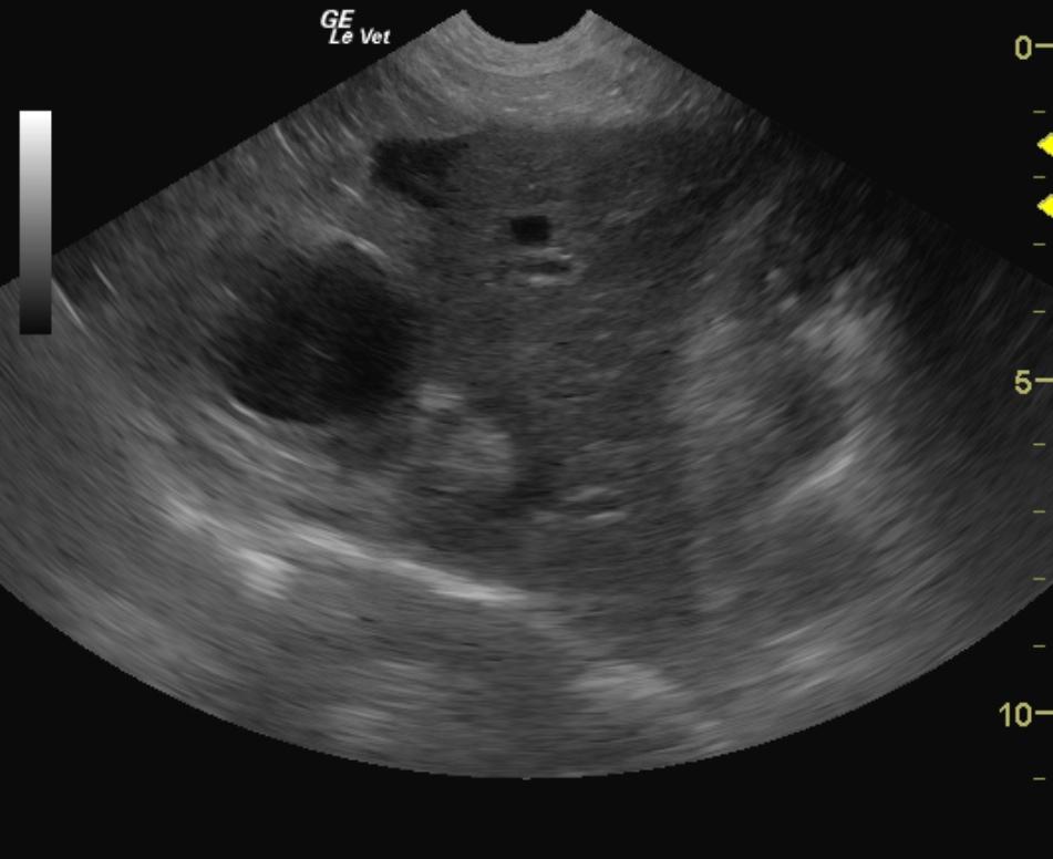
The abdomen in this patient presented excessively small liver with increased portal markings and coarse parenchyma with irregular capsular changes consistent with chronic hepatic fibrosis, cholangiohepatitis and portal hypertension given the ascites present. The gallbladder was thickened and mildly distended. The hepatic veins were excessively dilated due to over circulation and aggressive fluid therapy, therefore biopsies were not performed. 12-14 hours of diminishing fluid rate should provide for a safer window for ultrasound guided biopsy to assess parenchymal changes and rule out underlying lymphoma or other neoplastic event as well as assess for other source of cholangiohepatitis such as copper storage disease. Some ascites was noted around the upper gastrointestinal tract adjacent to the liver lobe. The omentum was mildly echogenic due to emerging ascites. The kidneys presented subcapsular fluid accumulation with diffuse interstitial pattern. Chronic leptospirosis can also present in this manner.
DX
Outcome
Antibiotic therapy was changed from penicillin to ampicillin and steroids were added. Leptosporosis titer results were positive for L. pomona, L. icterohaemorrhagiae, L. canicola, L. grippotyphosa, and L. autumnalis. Preprandial bile acids were high. Blood chemistry showed hyperphosphatemia, elevated ALT activity, hyperbilirubinemia, hypokalemia, and slightly elevated creatinine. Baytril and doxycycline were added to treatment plan and fluid therapy was continued. 24 hours later she was eating small amounts of food. On physical examination the patient had a depressed attitude, the abdomen was tender on palpation, and the icterus persisted. Patient was treated with Actigal, Denosyl, steroids, doxycycline, and amoxicillin. Several days later the patient had improved, was eating better, and there was no visible icterus. Follow-up blood work 2 weeks later showed ongoing improvement in the liver values. Continuation of the Denosyl was advised.
Clinical Differential Diagnosis
Hemolytic pathology: infectious, immune mediated vaccine reaction. Liver pathology: acute infectious, toxic, vaccine reaction; gallbladder obstruction
Sampling
None taken
Video
Patient Information
Clinical Signs
- Diarrhea
- Vomiting
History
- Corneal ulcer
Exam Finding
- Icterus
Images
Clinical Signs
- Diarrhea
- Vomiting
Special Testing
- 4Dx Negative