11 year old MN Pit-bull was presented with clinical signs of colitis of several months’ duration which had been controlled with anti-inflammatory dosages of prednisone. An abdominal mass was palpated on physical exam. The CBC was within normal limits. A mild elevation of the ALP enzyme activity was present on the serum biochemical profile.
11 year old MN Pit-bull was presented with clinical signs of colitis of several months’ duration which had been controlled with anti-inflammatory dosages of prednisone. An abdominal mass was palpated on physical exam. The CBC was within normal limits. A mild elevation of the ALP enzyme activity was present on the serum biochemical profile.
Case Study
Cecal leiomyoma diagnosed on ex-lap, in an 11 year old MN Pit Bull dog
Sonographic Differential Diagnosis
The intestinal mass is suggestive of sarcoma or carcinoma. Granulomatous disease is considered less likely.
Image Interpretation
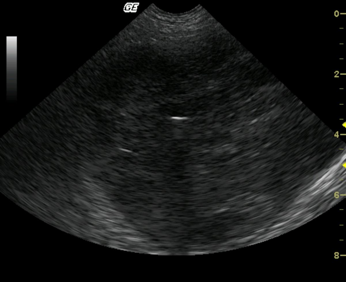
A large, dramatically hypoechoic, micronodular intestinal mass was present in the mid-abdomen. The lesion appeared isolated and therefore resectable. The linear hypoechoic lumen identifies this mass as intestinal in origin but the portion of intestine could not be identified due to complete loss of structural points of reference.
DX
Cecal leiomyoma
Outcome
The patient recovered uneventfully and was asymptomatic 3 months post surgery.
Clinical Differential Diagnosis
Neoplasia (lymphoma, leiomyoma, leiomyosarcoma, mast cell tumor, adenocarcinoma), foreign body, granuloma, abscess.
Sampling
An exploratory laparotomy revealed a spherical, irregular, firm cecal mass measuring 12 cm in diameter. Multiple omental adhesions were present, as well as an adhesion of the right middle liver lobe to the gastric fundus. Histopathology revealed a leiomyoma.
Video
Patient Information
Patient Name :
Thor D
Gender :
Male, Neutered
Species :
Canine
Type of Imaging : Ultrasound
Status :
Complete
Liz Wuz Here :
Yes
Code :
04_00183
Clinical Signs
- Diarrhea
History
- Colitis
Exam Finding
- Bradycardia
- Palpable mass
Images
Blood Chemistry
- Alkaline Phosphatase (SAP), High
Clinical Signs
- Diarrhea