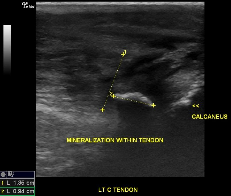
A 10-year-old MN Rottweiler mixed breed dog was presented with a history of limping on the left hind foot. Abnormalities on physical examination were pyrexia and a swollen left tarsus. A radiographic consultation found mild left stifle osteoarthrosis and joint effusion compatible with a partial or chronic cranial cruciate ligament rupture. Bursitis or partial avulsion of the common calcaneal tendon with enthesophyte formation was suspected. Lateral tarsal osteoarthrosis and mild coxofemeral osteoarthrosis was also noted.
A 10-year-old MN Rottweiler mixed breed dog was presented with a history of limping on the left hind foot. Abnormalities on physical examination were pyrexia and a swollen left tarsus. A radiographic consultation found mild left stifle osteoarthrosis and joint effusion compatible with a partial or chronic cranial cruciate ligament rupture. Bursitis or partial avulsion of the common calcaneal tendon with enthesophyte formation was suspected. Lateral tarsal osteoarthrosis and mild coxofemeral osteoarthrosis was also noted.