Bladder mass with renal dystrophy, suspect transitional cell carcinoma (TCC) in a 11 year old MN DSH cat

Case Study

Bladder mass with renal dystrophy, suspect transitional cell carcinoma (TCC) in a 11 year old MN DSH cat

This 11 year old MN DS cat has a history of hematuria/stranguria/pollakuria of 10 months duration. 

CBC/Chem: BUN 59, WBC low 2900

Urinalysis: USPG 1.016, protein +1, blood +3, WBCs 2-3/hpf, RBCs 21-50/hpf
This 11 year old MN DS cat has a history of hematuria/stranguria/pollakuria of 10 months duration. 

CBC/Chem: BUN 59, WBC low 2900

Urinalysis: USPG 1.016, protein +1, blood +3, WBCs 2-3/hpf, RBCs 21-50/hpf
DX

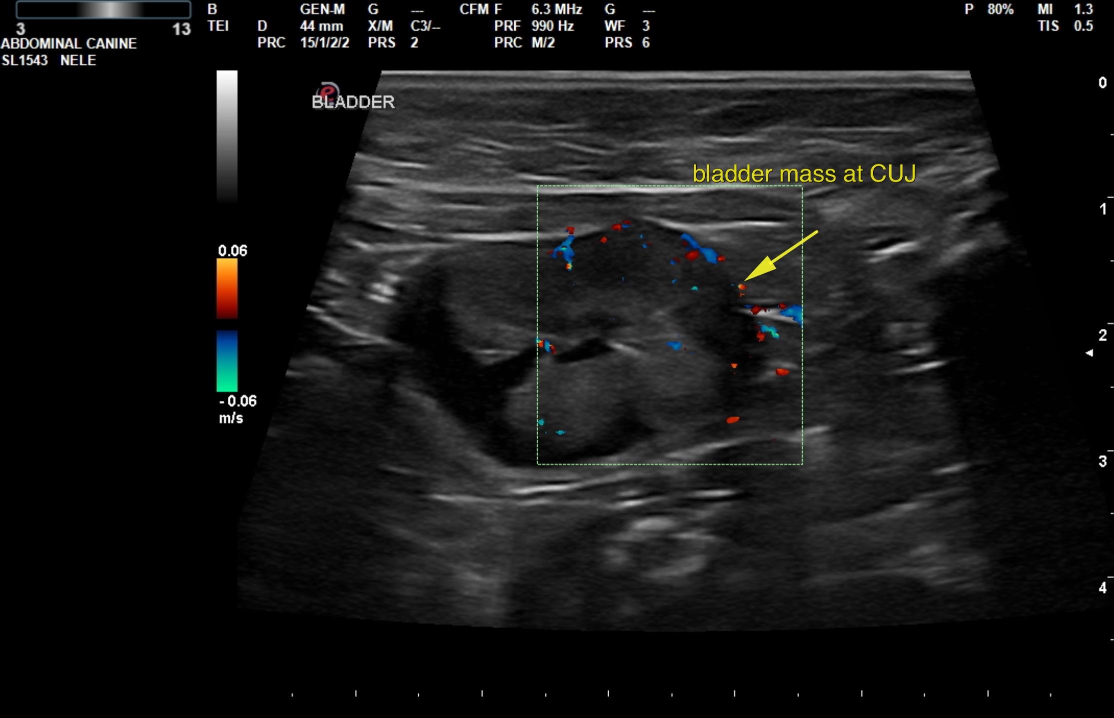
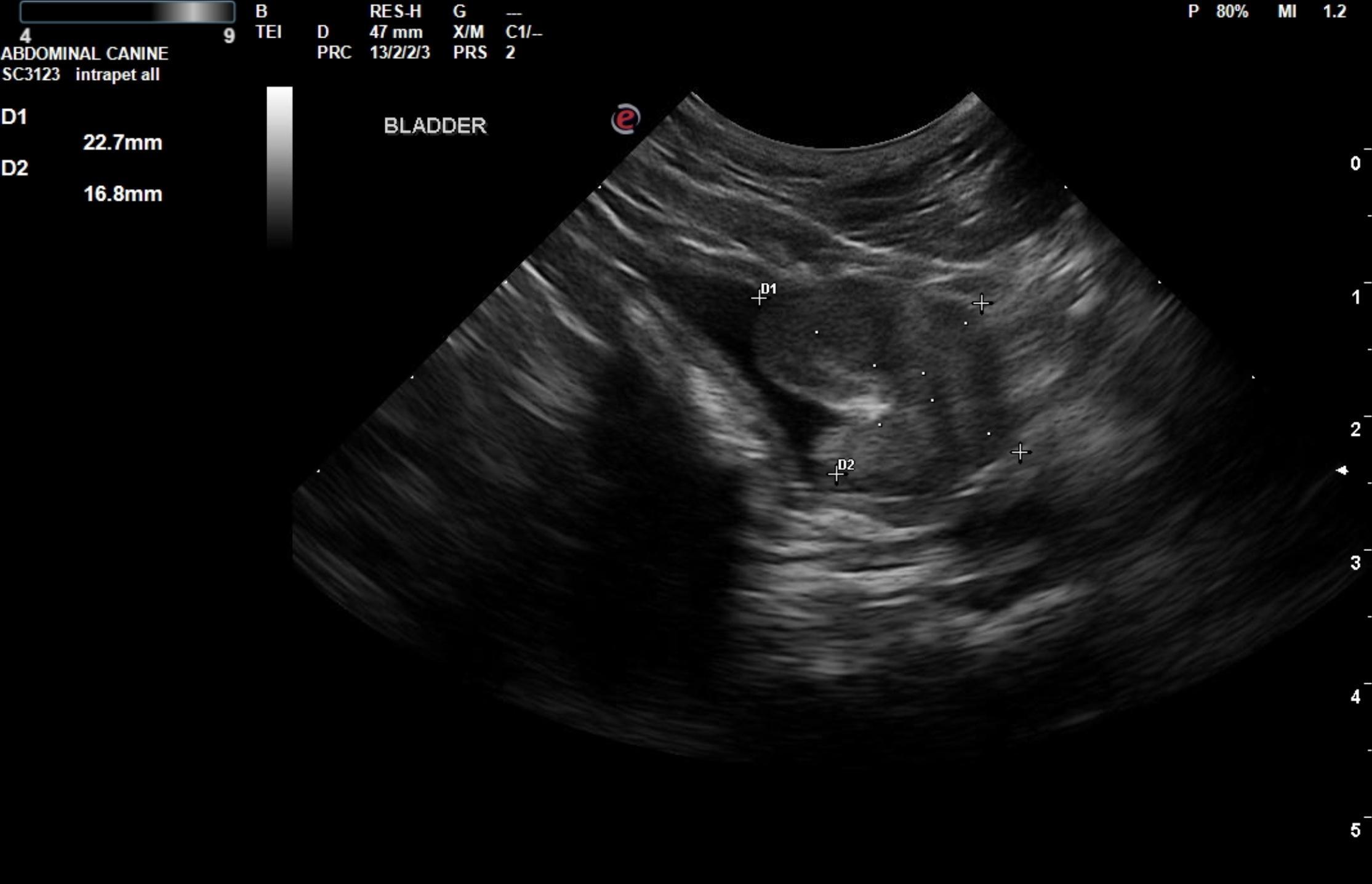
Bladder mass, focal mineralization suggestive for transitional cell carcinoma – potentially resectable. Renal dystrophy. Splenic and hepatic nodules; likely unrelated to the bladder mass. Suspect transitional cell carcinoma.

Image Interpretation


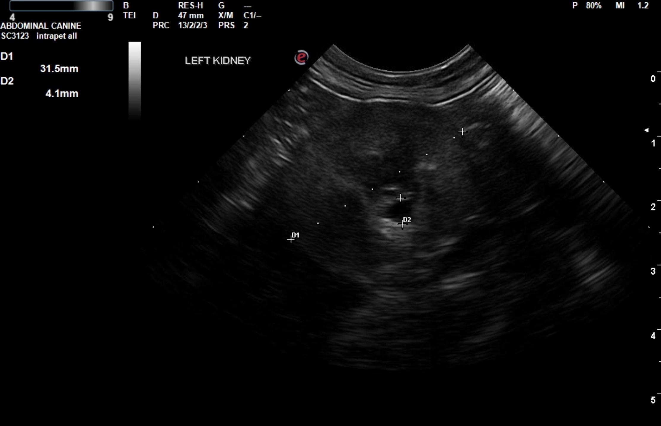
 The kidneys revealed largely normal size and structure, corticomedullary definition and ratio (cortex 1/3 of medulla) were essentially maintained with some age related loss of curvilinear patterns – mineralization, mild. The cortices presented largely uniform texture with some age related echogenic changes that are not likely of clinical significance at this time unless inflammatory sediment or proteinuria is present. Medullary echogenicity differed distinctly from that of the cortex and no evidence or dilation could be seen. The capsules were acceptably uniform for this age patient without dramatic irregularities. Pyelectasia of the left kidney was noted 0.41 cm. 


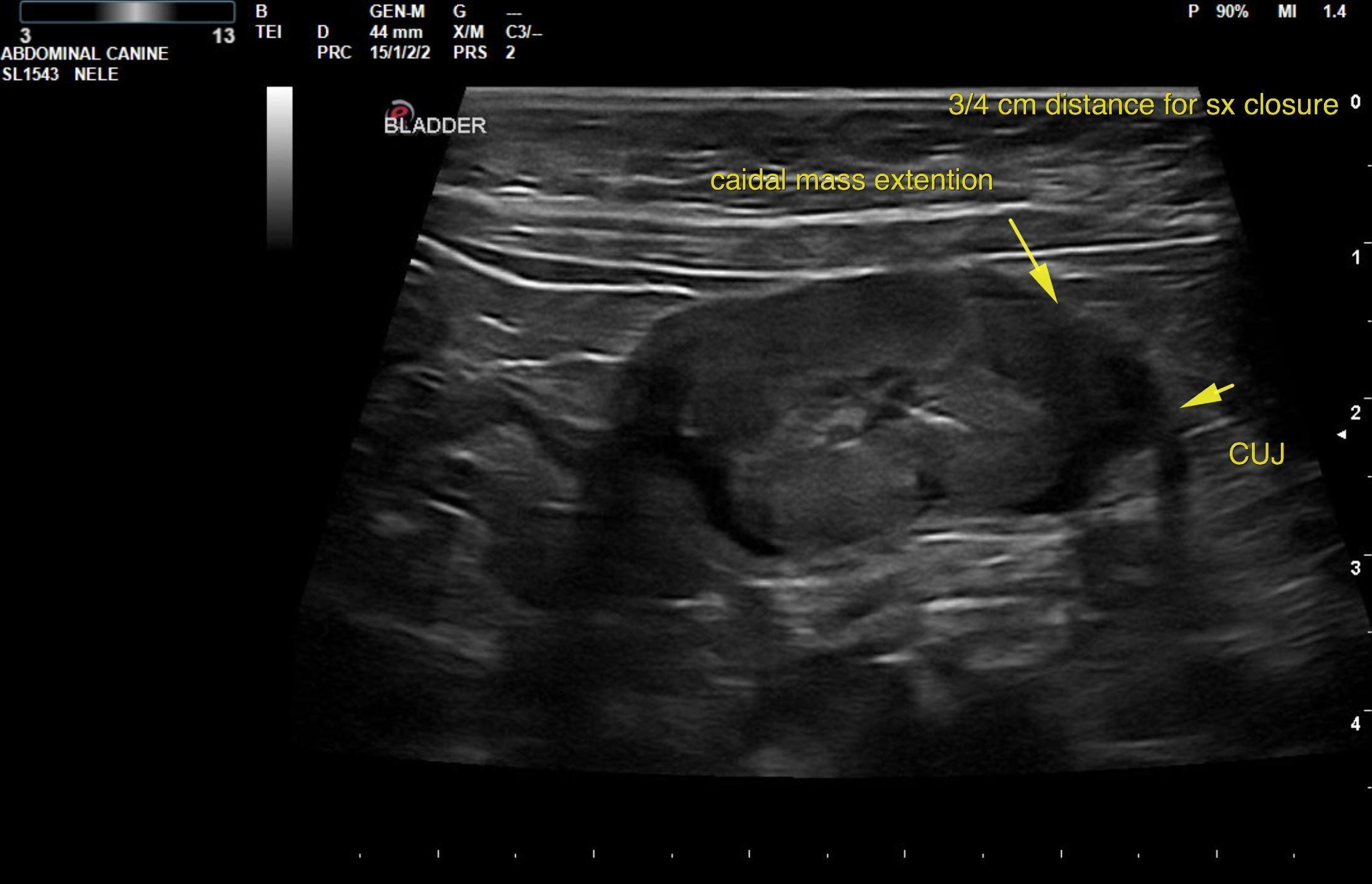
 Urinary bladder presented a concentric mass deriving from the cystourethral junction occupying the majority of the bladder. Moderate Doppler signals noted. Ureters were not obstructed. The mass appeared to be deriving from the ventral. Focal areas of mineralization were noted strongly suggestive for transitional cell carcinoma. The mass may be resectable with aggressive resection as approximately 0.75 cm of normal tissue between the caudal extension of the bladder mass and the cystourethral junction as noted. 

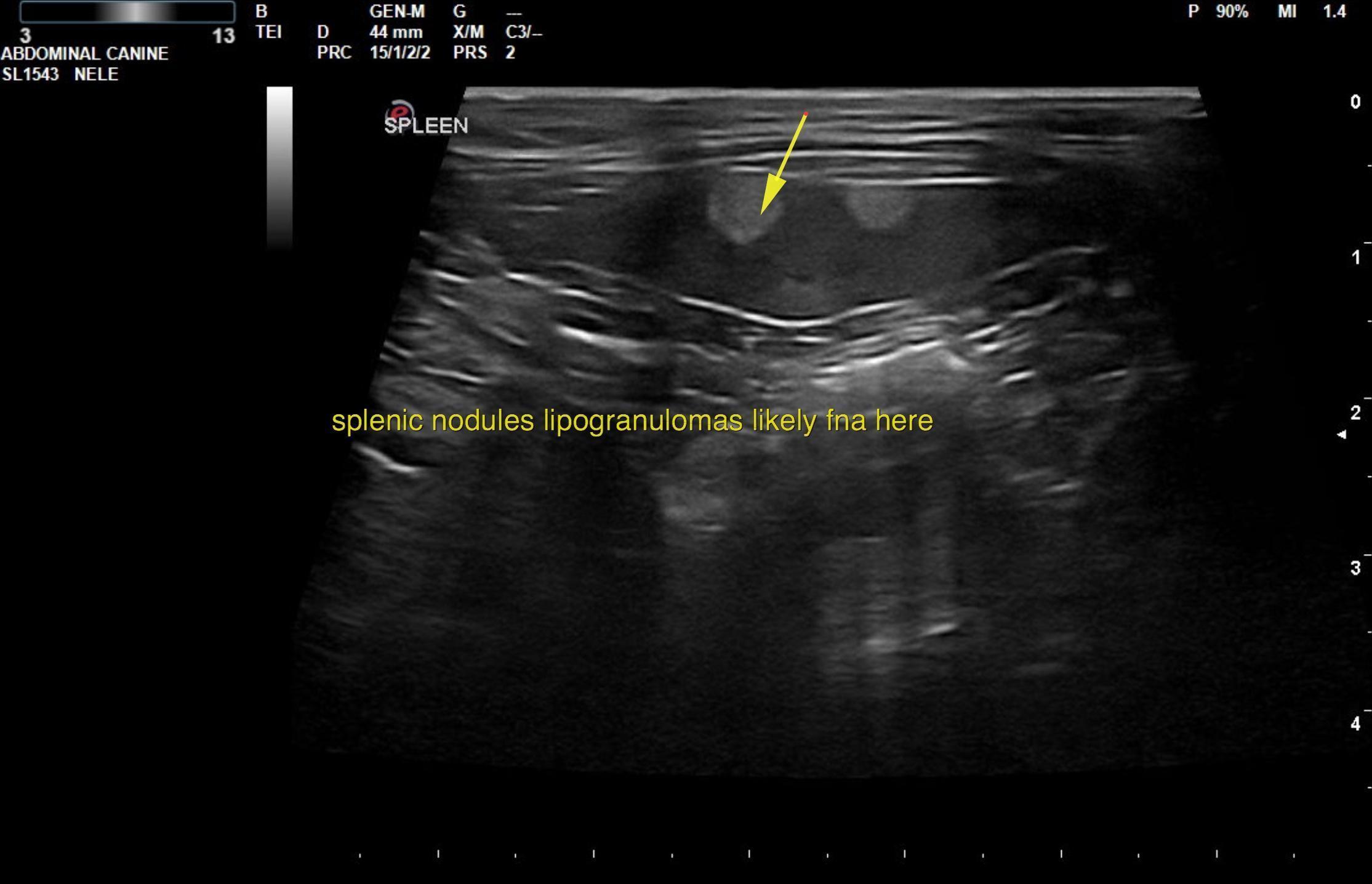
The spleen was largely normal, yet focal hyperechoic nodules were noted. 

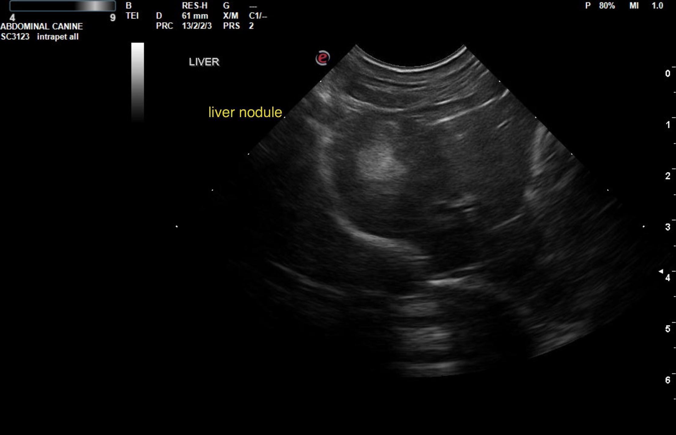
The liver revealed a hyperechoic nodule left cranial liver. 

The gastrointestinal presentation revealed mild uniform prominence of the gastric mucosa as well as areas of “ropey” small intestinal wall- minor. The muscularis layer was hypertrophied inverting the normal ratio (1:3). The intestinal mucosa was slightly irregular, thickened and hyperechoic suggestive of low grade, chronic inflammation. No evidence of obstruction was present. 

Outcome

FNA of the splenic nodules and liver nodule is recommended for screening in this patient followed by attempt at bladder mass resection by removal of the ventral bladder wall; however, closure may be difficult.
Complete ventral wall ablation may be the best option in this patient. Traumatic catheterization is recommended for definitive diagnosis

Comments

Chronic inflammatory bowel disease is probable with a low possibility of an early neoplastic event such as lymphoma. Full thickness tissue biopsies via open laparotomy would be necessary to rule out this possibility. 

Clinical Differential Diagnosis

renal pathology – cystitis vs TCC vs other

Patient Information

Patient Name : Mr. Cat Grosedale
Gender : Male, Neutered
Species : Feline
Type of Imaging : Ultrasound
Status : Complete
Liz Wuz Here : Yes
Code : 06_00362

Clinical Signs

  • Hematuria
  • Pollakiuria
  • Stranguria

Images

rb1rb4rb6rb3rb5rb8

Blood Chemistry

  • BUN high

CBC

  • WBC, Low

Clinical Signs

  • Hematuria
  • Pollakiuria
  • Stranguria

Urinalysi

  • Blood Present
  • Protein Present
  • Specific Gravity Low
  • WBCs Present
Skip to content