Bladder mass occupying trigone, CUJ and majority of bladder, likely TCC, in a 14 year old MN Australian Shepherd

Case Study

Bladder mass occupying trigone, CUJ and majority of bladder, likely TCC, in a 14 year old MN Australian Shepherd

A 14-year-old MN Australian Shepherd was presented for evaluation of a 10-day duration of hematuria, urinary incontinence, and PU/PD that had not improved with enrofloxacin. CBC was within reference range and abnormalities on serum biochemistry were mildly elevated ALP activity and cholesterol. Survey radiographs showed irregular mineralization in the trigone area of the bladder. 

A 14-year-old MN Australian Shepherd was presented for evaluation of a 10-day duration of hematuria, urinary incontinence, and PU/PD that had not improved with enrofloxacin. CBC was within reference range and abnormalities on serum biochemistry were mildly elevated ALP activity and cholesterol. Survey radiographs showed irregular mineralization in the trigone area of the bladder. 

DX

Bladder mass, not likely resectable. Possible early micrometastasis to the urethra or passage of small calculi.

Image Interpretation

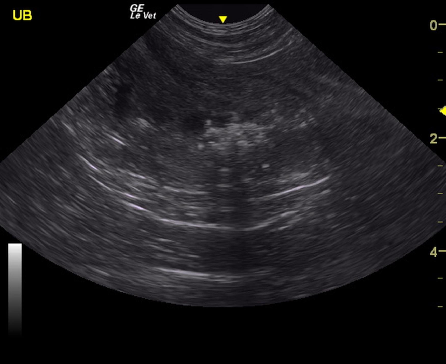
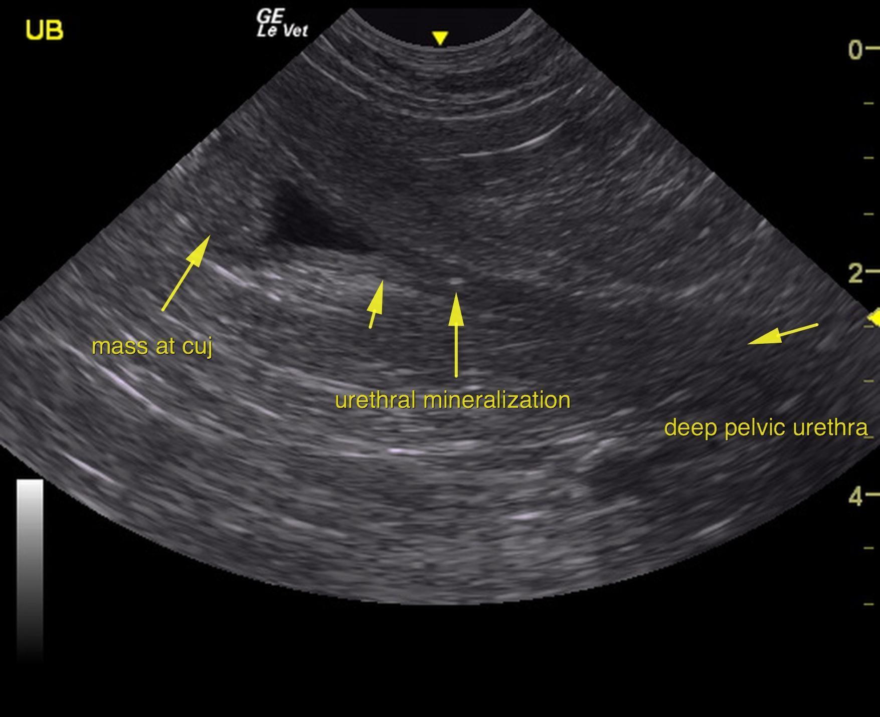
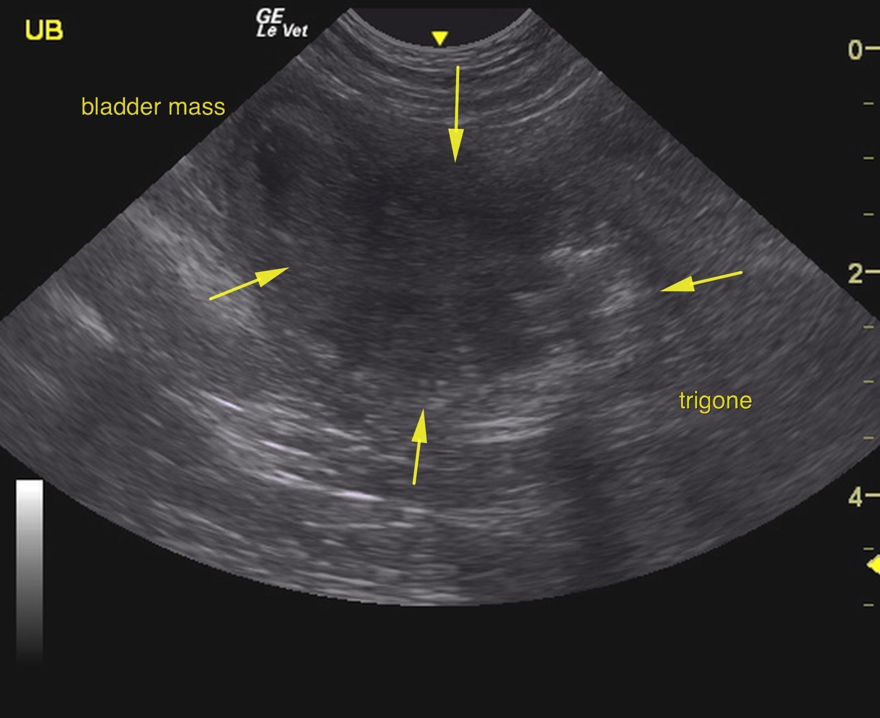
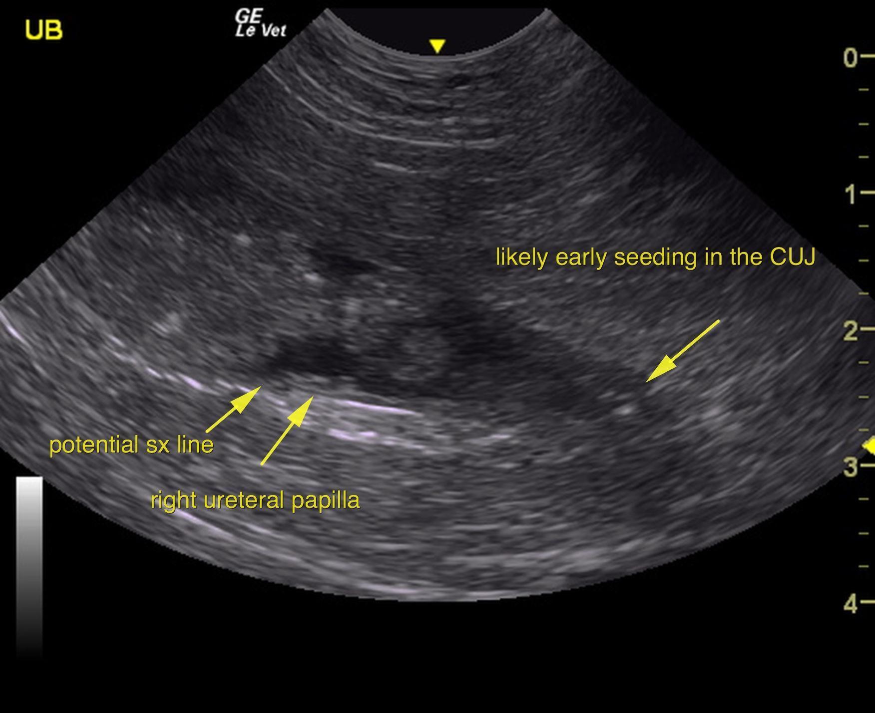
The urinary bladder presented a large, mineralizing mass that occupied the trigone and cystourethral junction as well as majority of the bladder. The mass measured approximately 4.0 x 3.0 cm and was moderately vascular. The mass appeared to be largely deriving from the ventral wall.  Mineralizing pattern is consistent with transitional cell carcinoma. The mass does not appear resectable. Kissing lesion polyps were noted in the dorsal wall as well. Free calculi were also noted. The iliac trifurcation was unremarkable with no evidence of pathology or metastatic disease. Slight mineralization was noted in the urethra. Potential passing calculi or early seeding is possible. The pelvic urethra was imaged 3.0 cm beyond the cystourethral junction. Minor tissue thickening at the cystourethral junction may represent seeding caudally not allowing for complete surgical resection. The bladder reaches a point of 0.5 cm from the right ureteral papillae leaving minimal amount of space for surgical closure. 

closure.

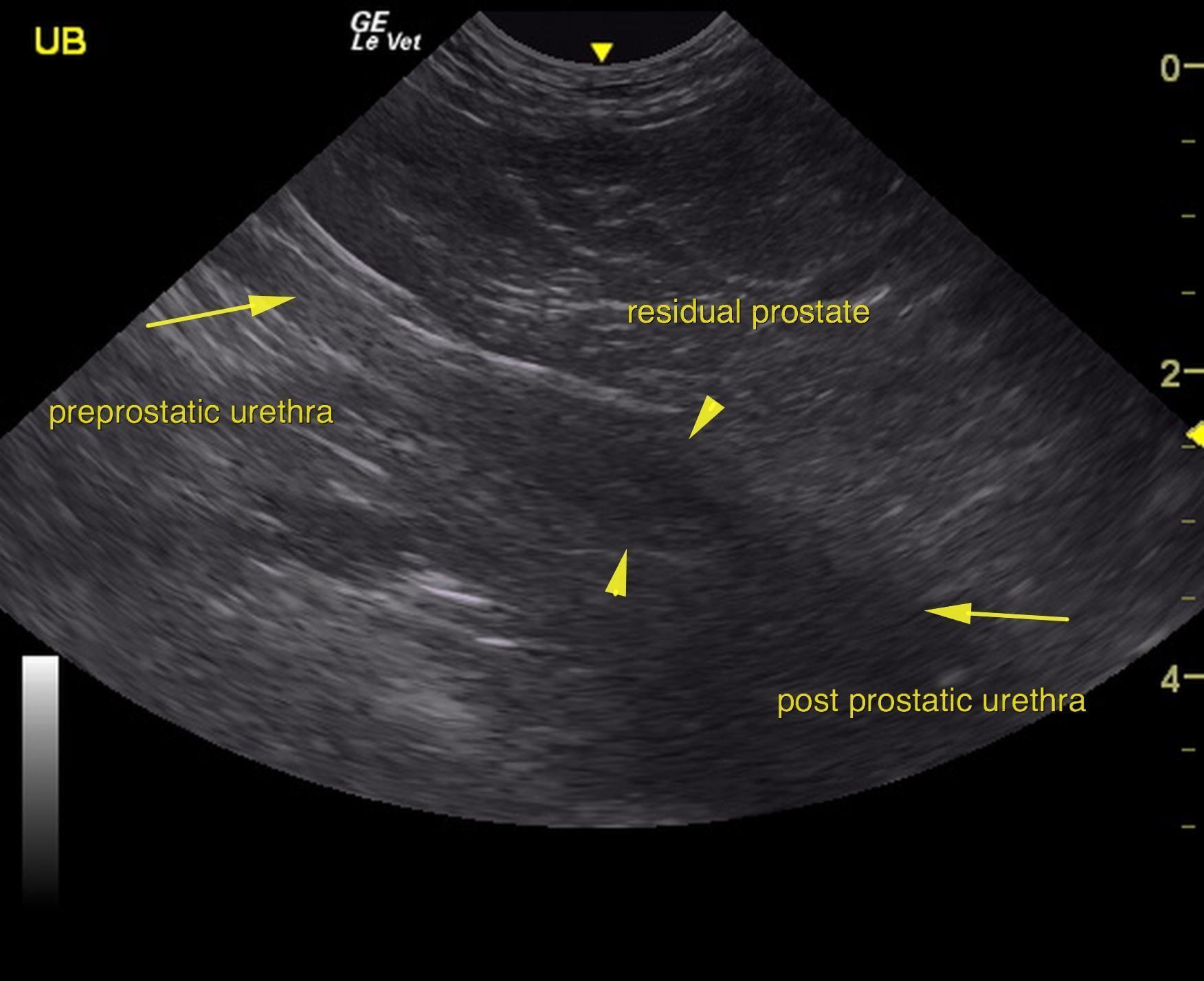
The residual prostate was uniform with no obvious seeding of the tumor.

The kidneys revealed largely normal size and structure, corticomedullary definition and ratio (cortex 1/3 of medulla) were essentially maintained with some age related loss of curvilinear patterns. The cortices presented largely uniform texture with some age related echogenic changes that are not likely of clinical significance at this time unless inflammatory sediment or proteinuria is present. Medullary echogenicity differed distinctly from that of the cortex and no evidence or dilation could be seen. The capsules were acceptably uniform for this age patient without dramatic irregularities. The right kidney measured 6.36 cm. The left kidney measured 5.2 cm. 

Outcome

Surgical debulking with stent placement could be considered. However, the amount of tissue to close the bladder will be minimal. However, the benefit of applying potential palliative ureteral and urethral stents could also be considered in this particular case. Otherwise, palliative therapy for transitional cell carcinoma after traumatic catheterization confirms TCC could be considered. The best option in this patient to avoid abdominal seeding is to performed ultrasound-guided traumatic catheterization of the tumor.

Clinical Differential Diagnosis

Bladder – neoplasia, urolith
Prostate – neoplasia, abscess

Patient Information

Patient Name : Bojangles Gordon
Gender : Male, Neutered
Species : Canine
Type of Imaging : Ultrasound
Status : Complete
Liz Wuz Here : Yes
Code : 06_00369

Clinical Signs

  • Hematuria
  • Incontinence
  • PU-PD

Images

as7as8as9as6as11

Blood Chemistry

  • Alkaline Phosphatase (SAP), High
  • Cholesterol, Low

Clinical Signs

  • Hematuria
  • Incontinence
  • PU-PD
Skip to content