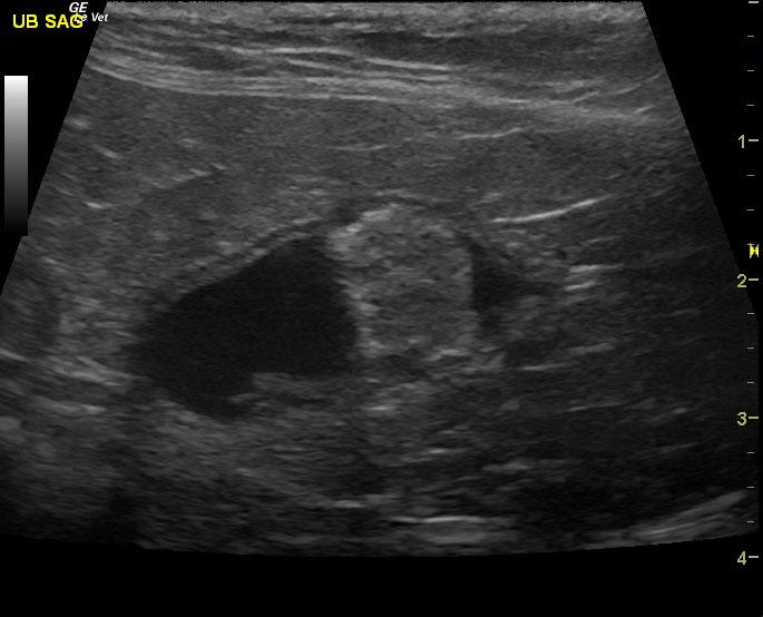
A 16-year-old neutered male DSH cat with a history of weight loss and inflammatory bowel disease was presented for poor appetite. Dehydration, dental disease, and nasal discharge were noted on physical examination. Hematuria was present on urinalysis. Initial CBC showed leukocytosis and anemia, which remained fairly unchanged after a month of antibiotic therapy.
A 16-year-old neutered male DSH cat with a history of weight loss and inflammatory bowel disease was presented for poor appetite. Dehydration, dental disease, and nasal discharge were noted on physical examination. Hematuria was present on urinalysis. Initial CBC showed leukocytosis and anemia, which remained fairly unchanged after a month of antibiotic therapy.