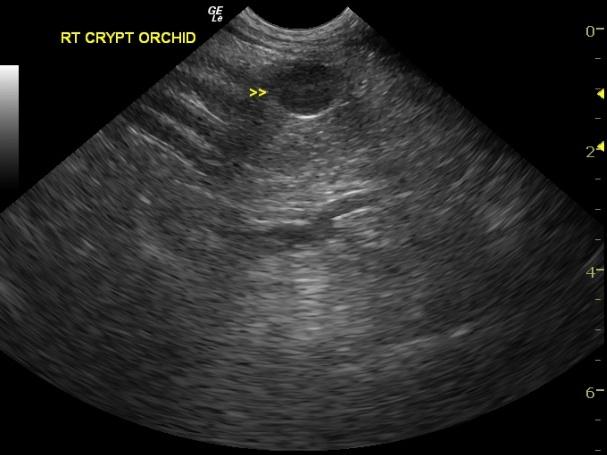
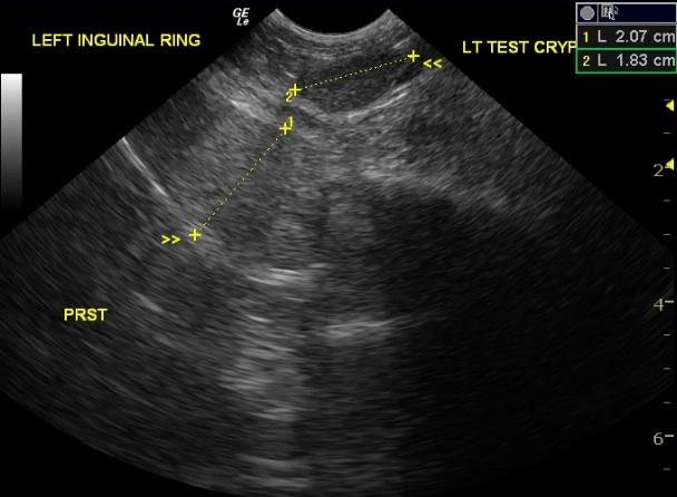
A 14 year old male Poodle presented for history of possible retained testicle. Clinical exam revealed an empty scrotal sac and lack of typical incision scar from neuter surgery. Rectal palpation revealed a prominent prostate.
A 14 year old male Poodle presented for history of possible retained testicle. Clinical exam revealed an empty scrotal sac and lack of typical incision scar from neuter surgery. Rectal palpation revealed a prominent prostate.