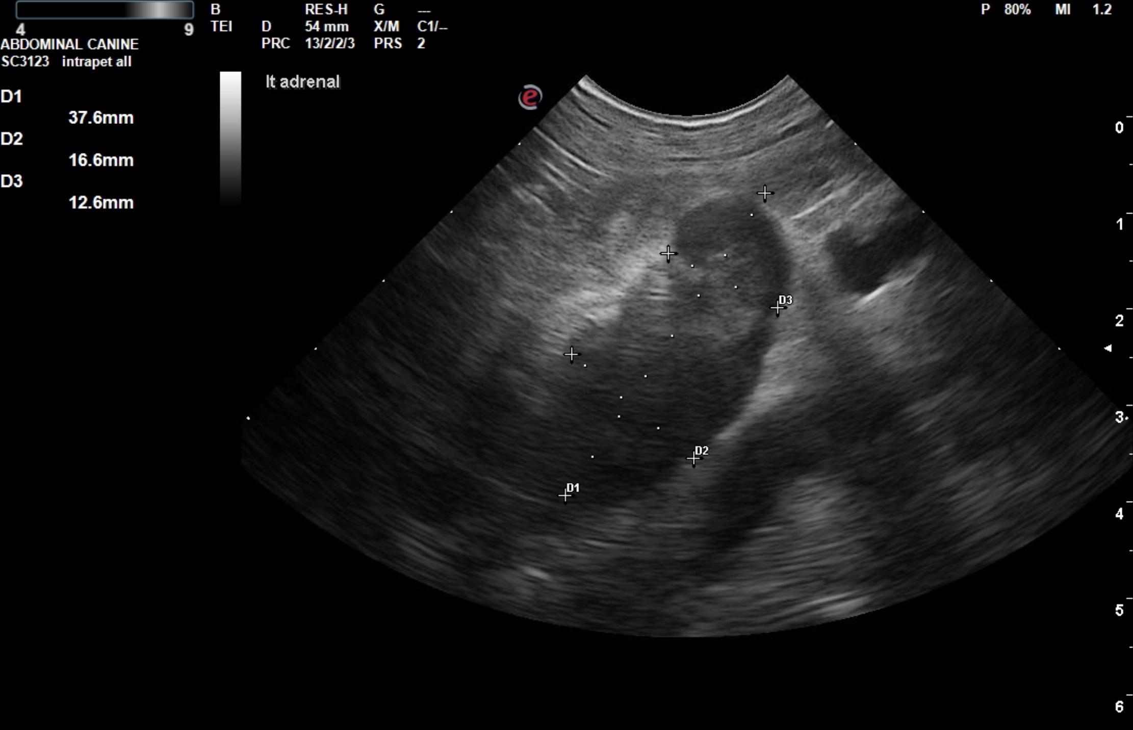
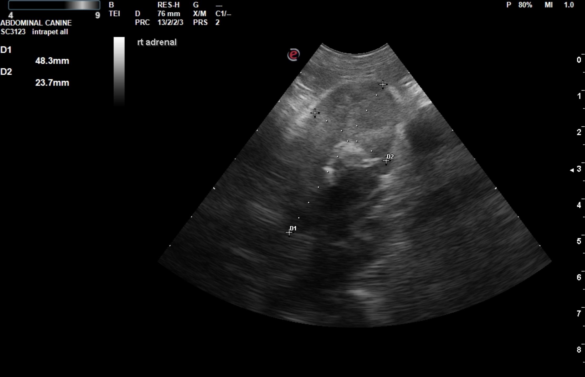
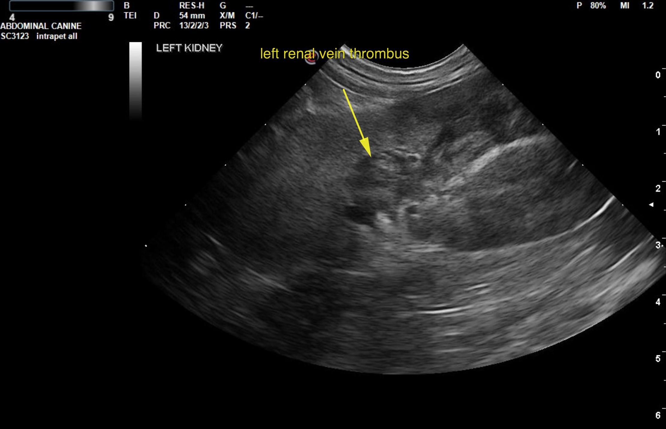
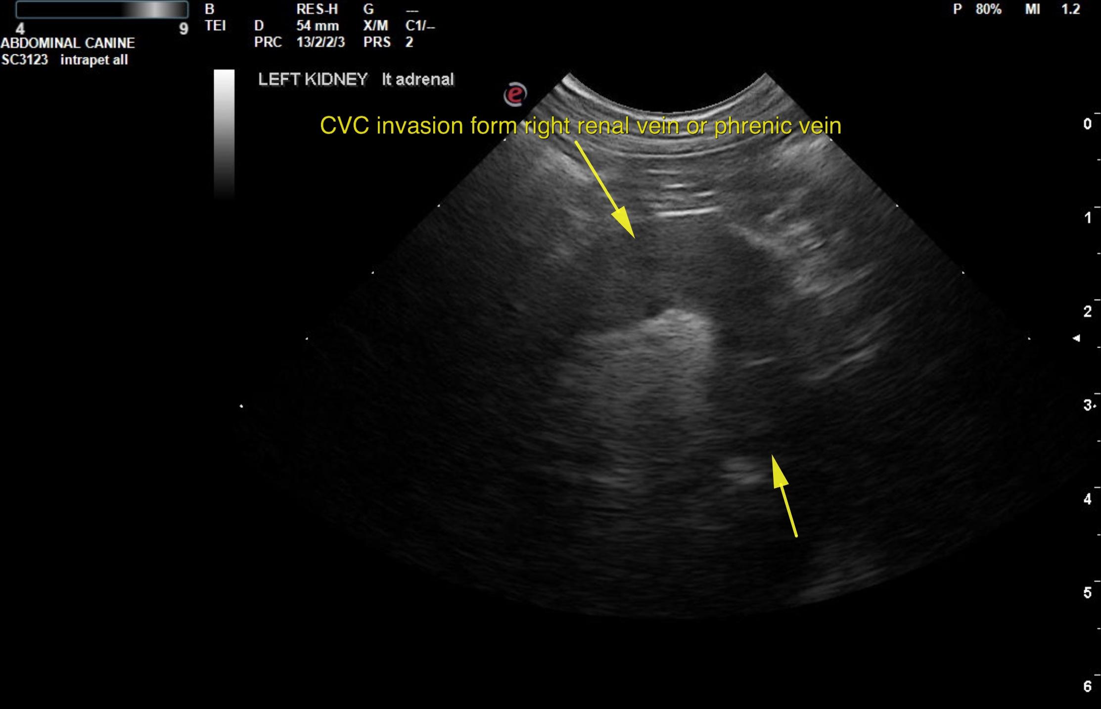
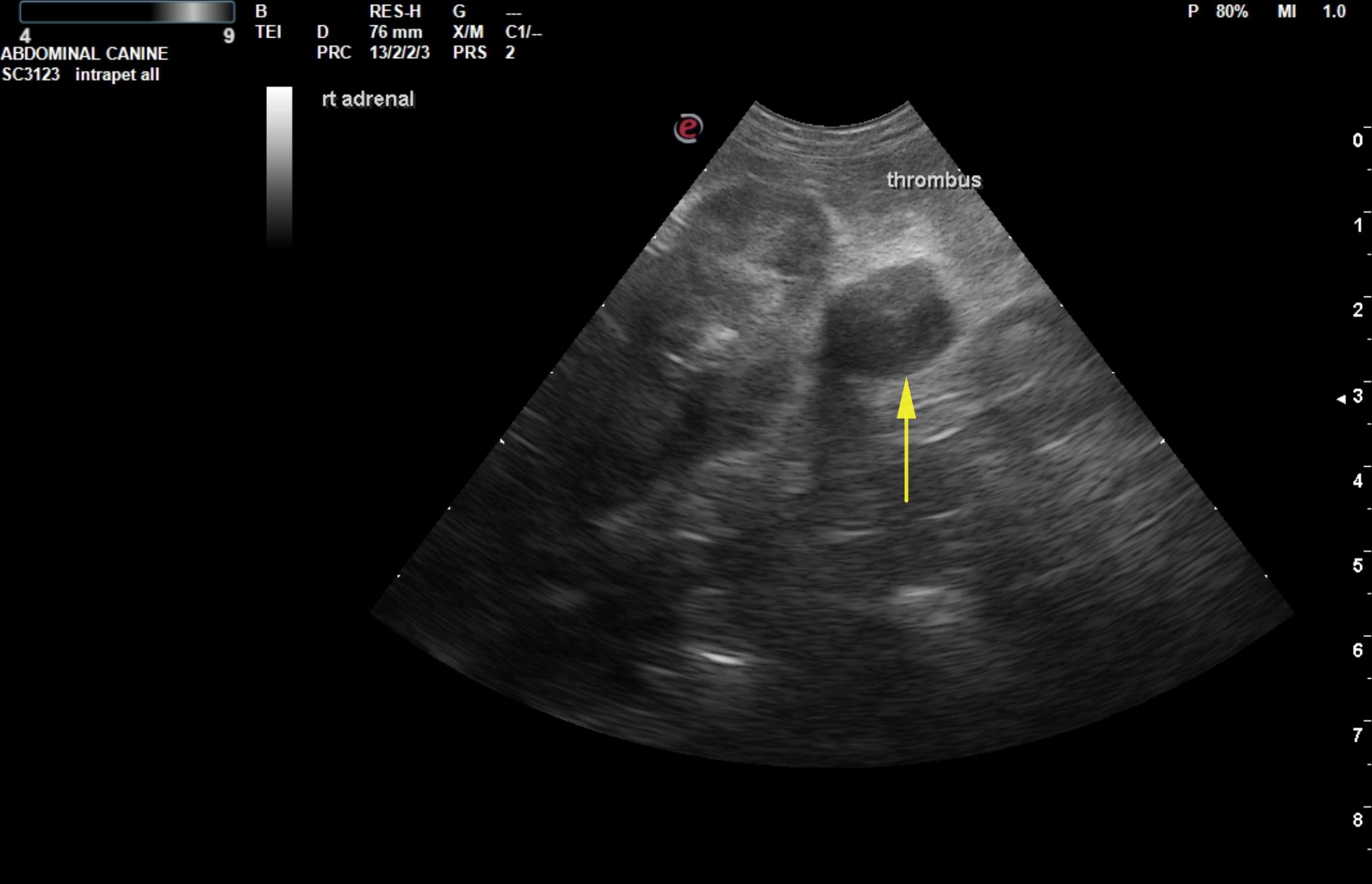
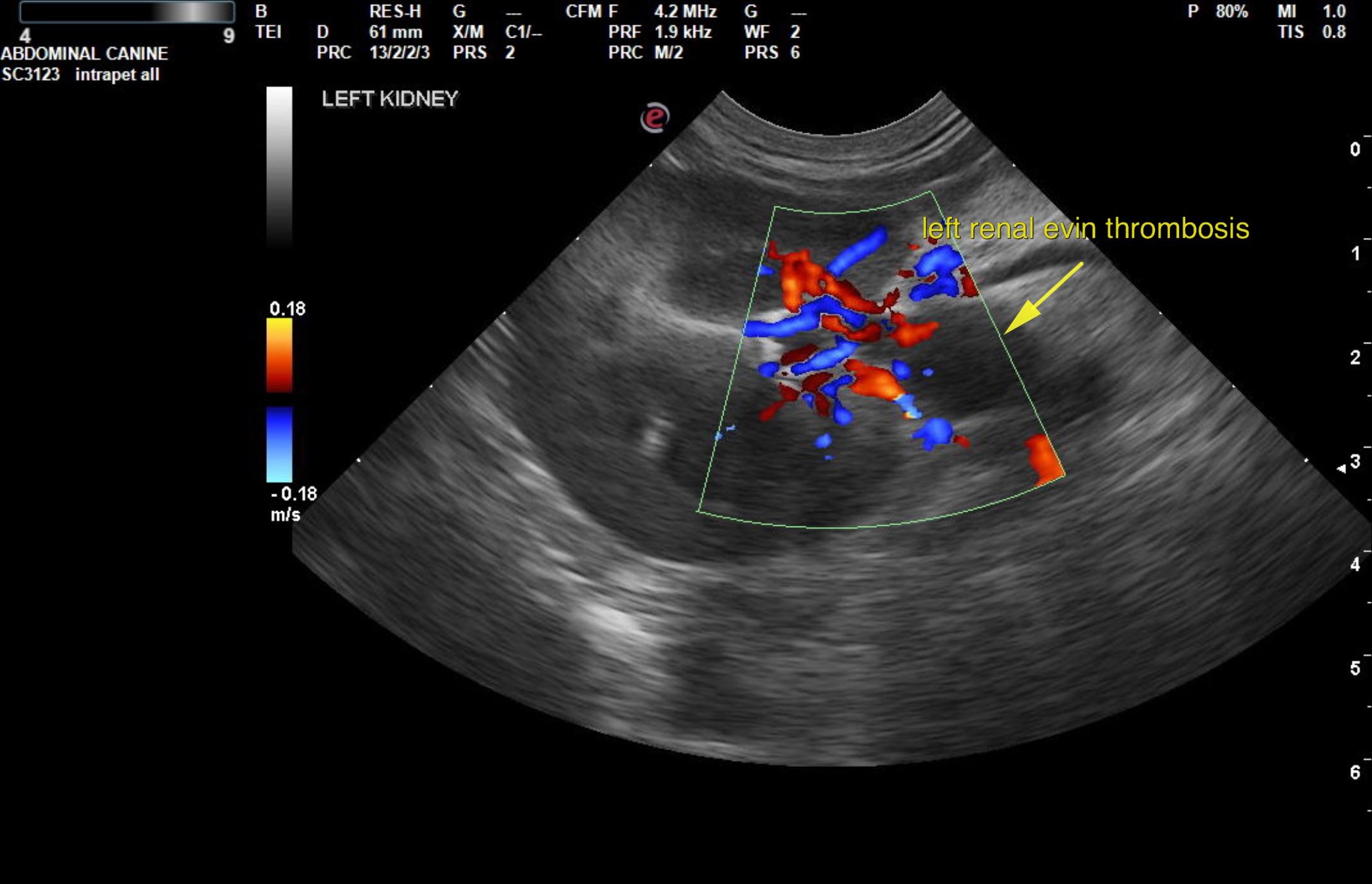
This 10 year old MN Cocker Spaniel dog presented with acute onset PU/PD. Recent anorexia, cranial abomen full on palpation and loss of detail near kidneyon rads. Glucose low.
This 10 year old MN Cocker Spaniel dog presented with acute onset PU/PD. Recent anorexia, cranial abomen full on palpation and loss of detail near kidneyon rads. Glucose low.