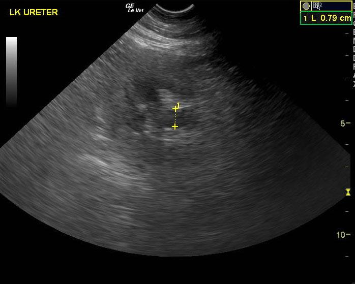
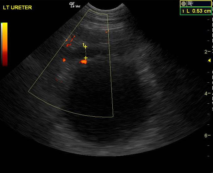
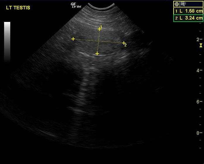
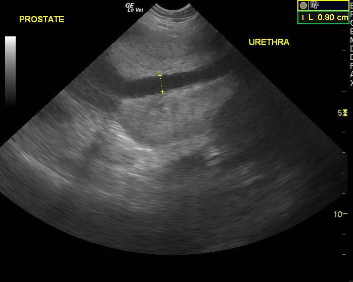
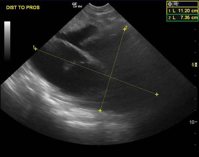
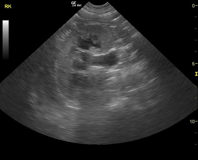
A 7 year old male intact German Shepherd Dog was presented for evaluation due to not defecating for the past few days, dribbling urine, and exhibiting lethargy. The patient was also cryptorchid, but the testicles had not been found surgically. On rectal palpation, digital entrance to the rectum was obstructed. UA and CBC/Chem were not available at the time of the sonogram.
A 7 year old male intact German Shepherd Dog was presented for evaluation due to not defecating for the past few days, dribbling urine, and exhibiting lethargy. The patient was also cryptorchid, but the testicles had not been found surgically. On rectal palpation, digital entrance to the rectum was obstructed. UA and CBC/Chem were not available at the time of the sonogram.