Adrenal mass in a 12 year old FS Yorkshire Terrier with sudden onset aggression

Case Study

Adrenal mass in a 12 year old FS Yorkshire Terrier with sudden onset aggression

A 12-year-old FS Yorkshire terrier dog was presented for sudden onset aggression. Two years previously, calcium oxalate uroliths had been removed via cystotomy. The dog was on Hill’s U/D and enacard. An inappropriate specific gravity, 3+ protein, hematuria, and elevated urine protein: creatinine ratio was present on urinalysis. CBC was within normal limits, whereas serum biochemistry showed elevated ALT and elevated ALP activity, hypercalcaemia, and elevated lipase. T4 was low.
A 12-year-old FS Yorkshire terrier dog was presented for sudden onset aggression. Two years previously, calcium oxalate uroliths had been removed via cystotomy. The dog was on Hill’s U/D and enacard. An inappropriate specific gravity, 3+ protein, hematuria, and elevated urine protein: creatinine ratio was present on urinalysis. CBC was within normal limits, whereas serum biochemistry showed elevated ALT and elevated ALP activity, hypercalcaemia, and elevated lipase. T4 was low.
DX

Right adrenal mass, renal calculi, biliary calculi, and CBD obstruction

Sonographic Differential Diagnosis

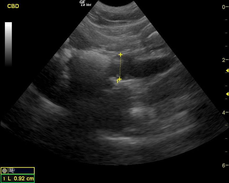
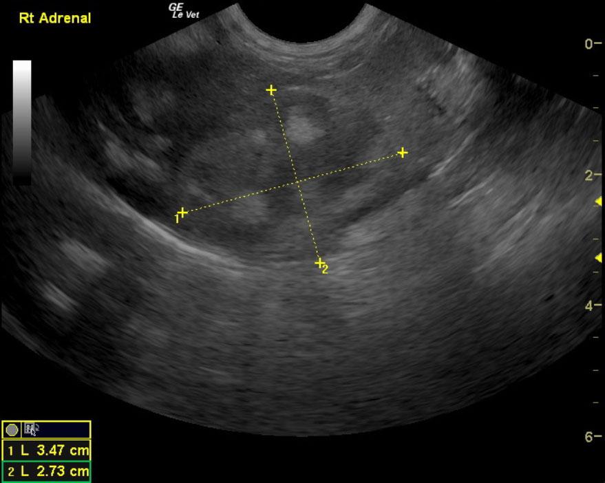
Common bile duct calculi and obstruction with an inflamed common bile duct and inspissated debris. There is a small calculus embedded at the duodenal papilla. Surgical intervention is highly recommended. Concurrent right adrenal gland mass, which is not invasive. Potential myelolipoma or benign hyperplasia. Other possibilities include pheochromocytoma or non functional adenocarcinoma given that PU/PD is not overtly present. Given that the urine specific gravity is persistently greater than 1.020, blood pressure measurements would be warranted. Renal fibrosis and calculi. Urine culture and sensitivity would be warranted as well as blood pressure measurements. Eventual surgical intervention to liberate the common bile duct would be recommended. It does appear to have adequate integrity. Therefore, incision into the cystic duct and manual manipulation of the calculus at the duodenal papilla and the larger calculi at the cystic duct enters into the common bile duct and should be adequate. There is the minor potential that cholecystoduodenostomy may be necessary. At the time of surgery a right adrenalectomy could also be performed. CT evaluation may be warranted. However, no overt evidence of invasion was noted in this patient. 25-gauge fine-needle aspirates of the right adrenal gland mass can also be considered with some possibility of complication. However, if surgical intervention for the common bile duct is necessary, then a right adrenalectomy would be ideal at that time.

Image Interpretation

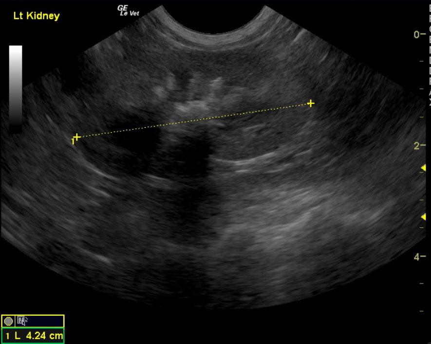
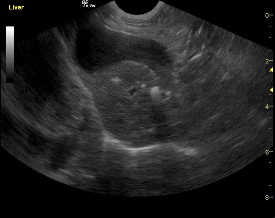
Both kidneys had a diffuse, interstitial nephrosis pattern with corticomedullary and medullary calculi. These calculi are likely calcium oxalates given the history and sonographic appearance. Occasional renal cortical cysts were noted. The left kidney measured 4.2 cm. The right kidney measured 4.4 cm. The left adrenal gland was normal in size and contour. However, the right adrenal gland comprised a mass that measured 3.5 x 2.7 cm with mixed, hyperechoic changes. This may represent a myelolipoma given that the patient is not persistently polyuric and polydipsic. Other possibilities are non-functional adenocarcinoma or pheochromocytoma as well as benign hyperplasia. This mass is expansive upon the right kidney, but no overt invasion into the vena cava was noted on these views. This appears resectable with some effort. Blood pressure monitoring would be recommended. The left adrenal gland measured 2 x 0.5 cm. The liver in this patient presented multiple lobar calculi with common bile duct calculi and significant dilation of the common bile duct. It measured 0.9 cm with echogenic debris and large calculus localized in the entrance of the cystic duct into the common bile duct. The distal aspect of the common bile duct also contained a calculus at the duodenal papilla that appears to be obstructing. The calculus at the entrance to the common bile duct measured 1.4 cm, but multiple other small calculi were noted. Around the common bile duct, there were some areas of periductile inflammation. The remainder of the liver presented minor, degenerative change; minor excessive gallbladder debris and distinction of the cystic duct was also noted.

Outcome

The patient was lost to follow up.

Clinical Differential Diagnosis

Aggression – Neurological (GME, neoplasia, cyst, abscess, metabolic encephalopathy), behavioral (senility), pain (orthopedic, neoplasia, urolith), liver disease, renal disease. Hypercalcemia – neoplasia, renal disease, toxicity, granulomatous disease.

Sampling

None

Patient Information

Patient Name : Lady F
Gender : Female, Spayed
Species : Canine
Type of Imaging : Ultrasound
Status : Complete
Liz Wuz Here : Yes
Code : 07_00060

Clinical Signs

  • Aggression

History

  • Calcium Oxalate Stones
  • Cystotomy
  • Enalapril therapy

Images

07_00060_image_01_0630201108122307_00060_image_02_0630201108123407_00060_image_03_0630201108124407_00060_image_04_06302011081257

Blood Chemistry

  • Alkaline Phosphatase (SAP), High
  • ALT (SGPT), High
  • Calcium, High
  • Hypothyroidism
  • Lipase, High

Clinical Signs

  • Aggression

Urinalysi

  • Blood Present
  • Specific Gravity Abnormal
  • Urine Protein:Creatinine High
Skip to content