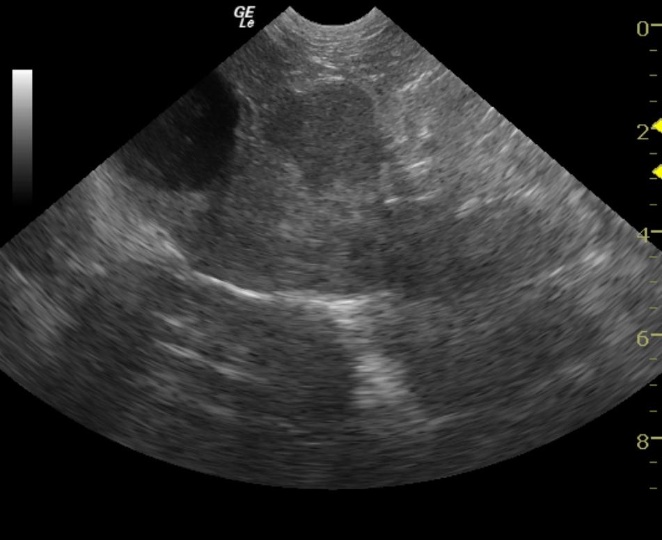
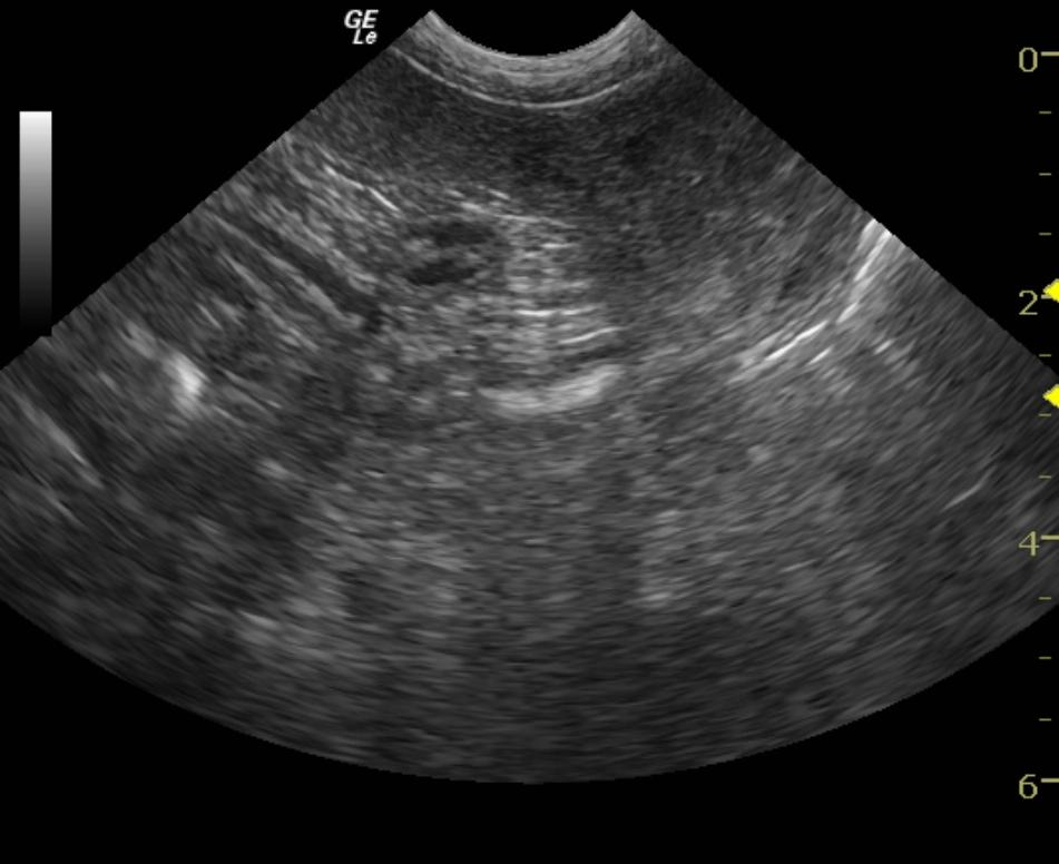
A 13 year old FS Miniature Schnauzer dog was presented due to clinical signs of recurrent urinary tract infections. CBC found a decreased hemoglobin and a high platelet count. In-house urinalysis showed a low specific gravity, high WBCs, high RBCs, and the presence of mucous strands. Blood chemistry revealed an increased ALT enzyme activity, hyperphosphatemia, elevated urea, elevated cholesterol, and elevated triglycerides.
A 13 year old FS Miniature Schnauzer dog was presented due to clinical signs of recurrent urinary tract infections. CBC found a decreased hemoglobin and a high platelet count. In-house urinalysis showed a low specific gravity, high WBCs, high RBCs, and the presence of mucous strands. Blood chemistry revealed an increased ALT enzyme activity, hyperphosphatemia, elevated urea, elevated cholesterol, and elevated triglycerides.