A 12-year-old MN Siamese cat was presented for evaluation of either syncopal episodes or seizures. On physical examination, arrhythmia and bradycardia were noted, with a HR that fluctuated from 80 to 160 BPM. Systolic BP was normal.
A 12-year-old MN Siamese cat was presented for evaluation of either syncopal episodes or seizures. On physical examination, arrhythmia and bradycardia were noted, with a HR that fluctuated from 80 to 160 BPM. Systolic BP was normal.
Case Study
15_00081 Minko S Narrow LVOT/AO
Sonographic Differential Diagnosis
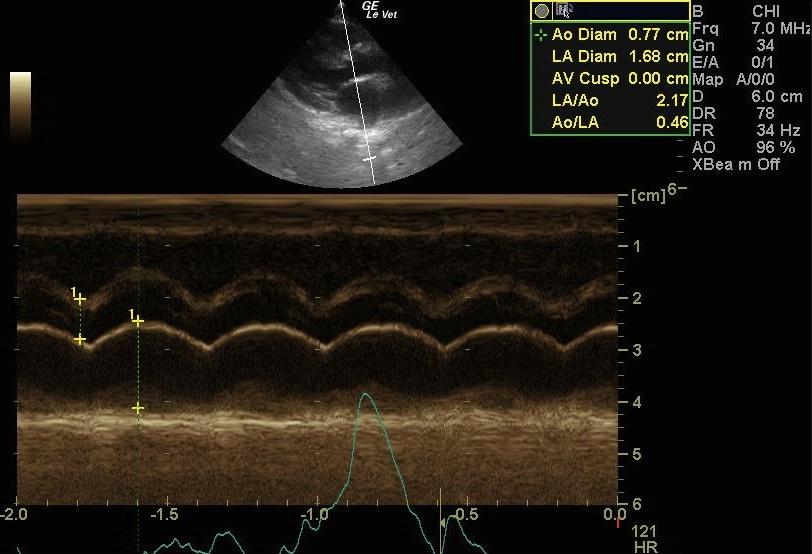
The enlarged left atrium (mild enlargement) may be either a cause of mild regurgitation (which could also be due to SAM) or a consequence of restriction to ventricular filling. The morphology and function of the heart (on echo) do not give any cause for the clinical problems.
Image Interpretation
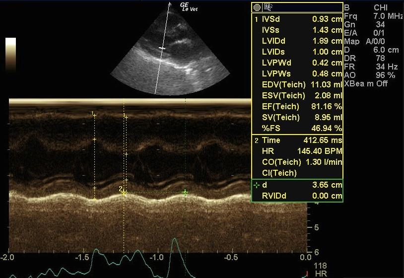
Normal left and right ventricle, an enlarged left atrium, and a slightly subnormal LVOT and AO diameter. CDI shows some slight mitral regurgitation. There is also possibly mild SAM but this cannot be determined on the 2D-clips. The AV occasionally flutters in systole; this may be due to turbulence within the LVOT. The concomitant ECG shows marked arrhythmia but the Sono-ECG is non-diagnostic.
DX
Normal left ventricle, enlarged left atrium, narrow LVOT/AO not of clinical importance.
Outcome
A Holter recording is strongly recommended in order to define the type and extent of the arrhythmia, as well as if the syncopal episodes occur during bradycardia, tachycardia or both. If this is not possible for the owner, a long ECG strip, with at least 3 leads, may help determine whether medication is indicated.
Comments
No further outcome provided.
Clinical Differential Diagnosis
Bradycardia – primary conduction anomaly, vagal-induced bradycardia, myocarditis, organophosphate toxicity, intra-cranial disease. Seizures – intra-cranial disease, infectious, metabolic, toxic, idiopathic.
Sampling
None
Video
Patient Information
Gender :
Male, Neutered
Species :
Feline
Type of Imaging : Ultrasound
Status :
Complete
Clinical Signs
- Seizures
- Syncopal episodes
Exam Finding
- Arrhythmia
- Bradycardia
- Fluctuating heart rate
Images
Clinical Signs
- Seizures
- Syncopal episodes