A 13-year-old FS DSH cat was presented for evaluation prior to a dental procedure. On physical examination a grade 3/6 murmur was present and blood pressure was normal.
A 13-year-old FS DSH cat was presented for evaluation prior to a dental procedure. On physical examination a grade 3/6 murmur was present and blood pressure was normal.
Case Study
15_00076 Barney K DRVOTO, SAM, HCM
Sonographic Differential Diagnosis
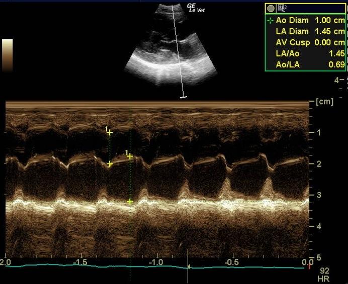
There are two sources for a heart murmur in this patient. While DRVOTO is considered normal in cats, SAM is clearly abnormal. The left ventricular hypertrophy could be due to HCM, but hyperthyroidism has to be excluded first.
Image Interpretation
Concentrically hypertrophied left ventricle (especially in the basal septal segment) and a slightly enlarged left atrium. The papillary muscles are hypertrophied. The mitral valve shows clear evidence of SAM (systolic anterior motion of the anterior leaflet). The flow profile across the LVOT appears dagger shaped, which is typical of dynamic obstruction of the left ventricular outflow tract (vmax 2.45 m/s, end systolic acceleration). The right ventricle and atrium are normal, but there is dynamic obstruction of the right ventricular outflow tract present (DRVOTO; indicated by the spectral Doppler flow profile).
DX
Hypertrophic cardiomyopathy with dynamic right ventricular outflow tract obstruction
Outcome
Since there is only minimal left atrial enlargement present, treatment is not indicated. Recheck every 6 months to evaluate the LA size would be recommended.
Comments
No further outcome provided.
Clinical Differential Diagnosis
Dilated/hypertrophic cardiomyopathy, vegetative endocarditis, mitral/tricuspid endocardiosis, ventricular hypertrophy secondary to hyperthyroidism, anemia.
Sampling
None
Video
Patient Information
Gender :
Female, Spayed
Species :
Feline
Type of Imaging : Ultrasound
Status :
Complete
Exam Finding
- Heart Murmur
Images