Skip to content
Customize Consent Preferences

We use cookies to help you navigate efficiently and perform certain functions. You will find detailed information about all cookies under each consent category below.

The cookies that are categorized as "Necessary" are stored on your browser as they are essential for enabling the basic functionalities of the site. ... 

Always Active

Necessary cookies are required to enable the basic features of this site, such as providing secure log-in or adjusting your consent preferences. These cookies do not store any personally identifiable data.

No cookies to display.

Functional cookies help perform certain functionalities like sharing the content of the website on social media platforms, collecting feedback, and other third-party features.

No cookies to display.

Analytical cookies are used to understand how visitors interact with the website. These cookies help provide information on metrics such as the number of visitors, bounce rate, traffic source, etc.

No cookies to display.

Performance cookies are used to understand and analyze the key performance indexes of the website which helps in delivering a better user experience for the visitors.

No cookies to display.

Advertisement cookies are used to provide visitors with customized advertisements based on the pages you visited previously and to analyze the effectiveness of the ad campaigns.

No cookies to display.

15-00222 Tilly E Subaortic stenosis and aortic regurg

Case Study

15-00222 Tilly E Subaortic stenosis and aortic regurg

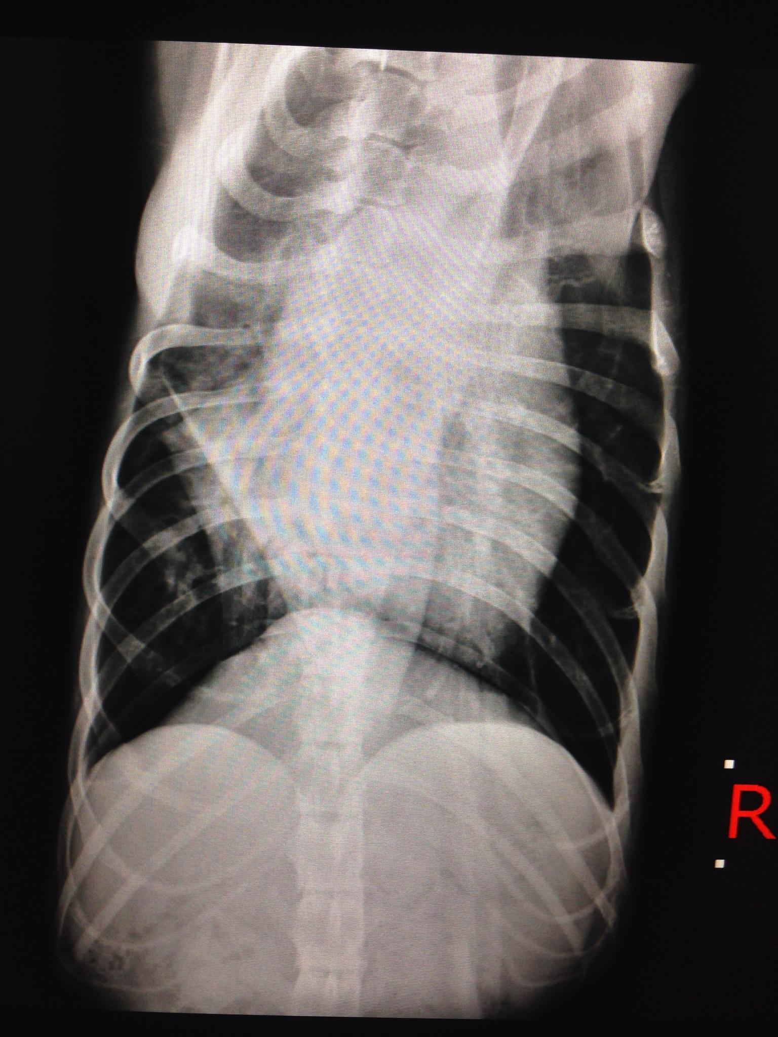
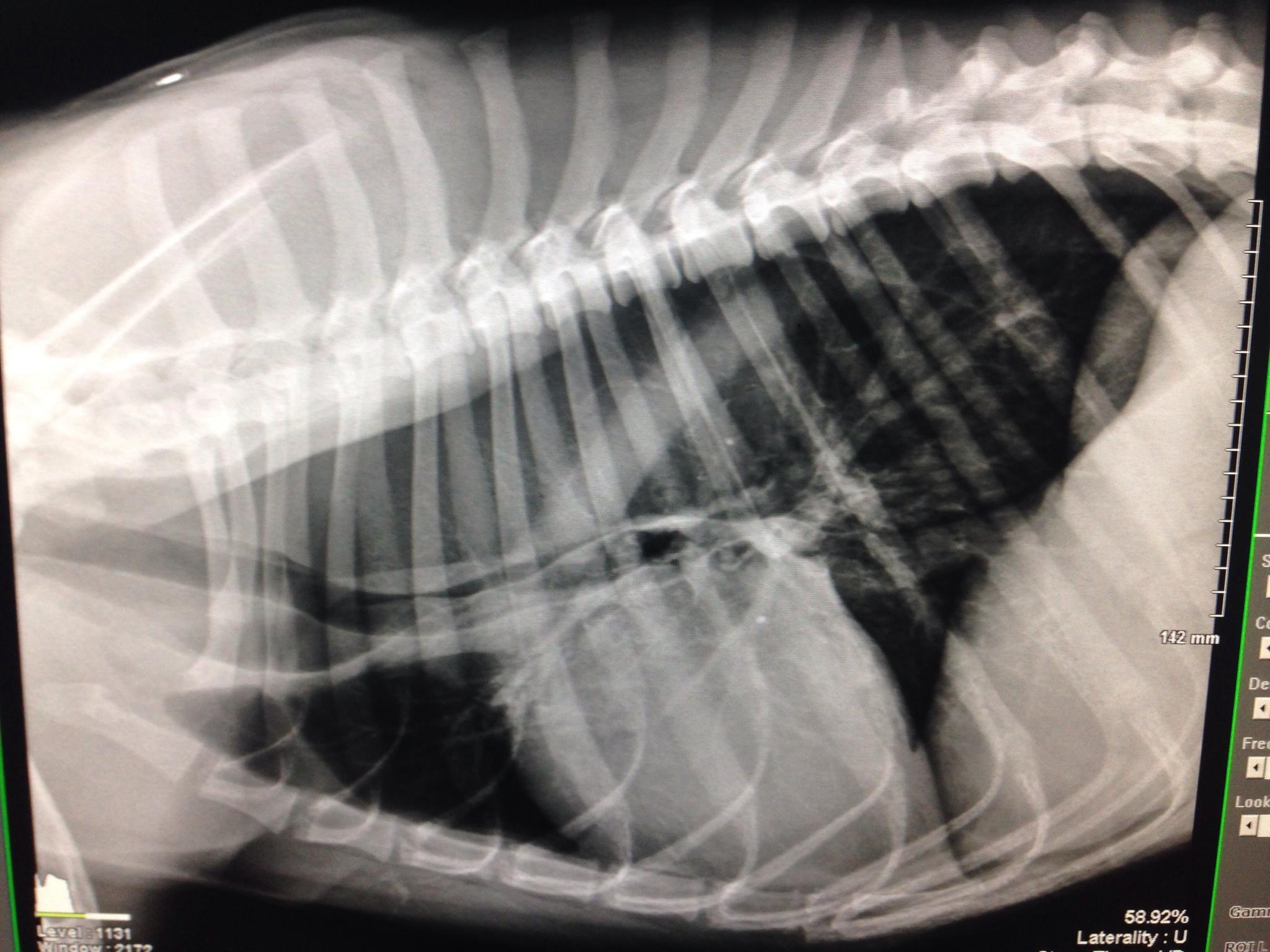
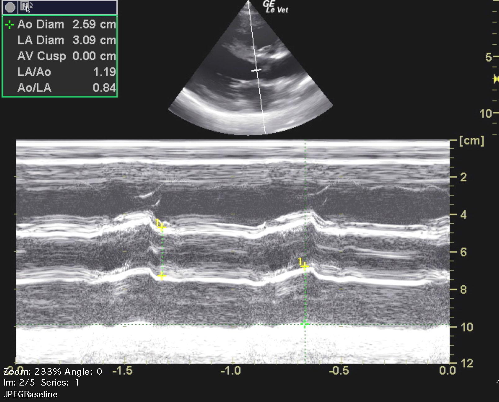
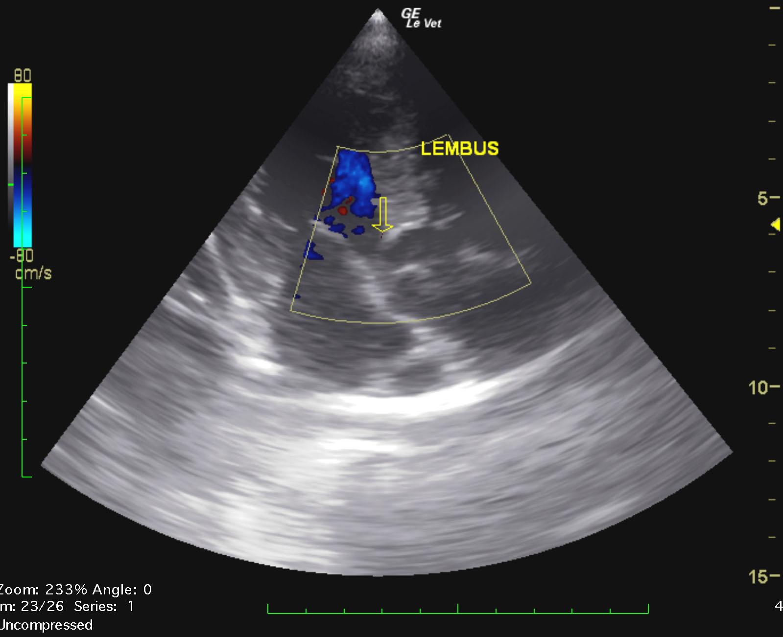
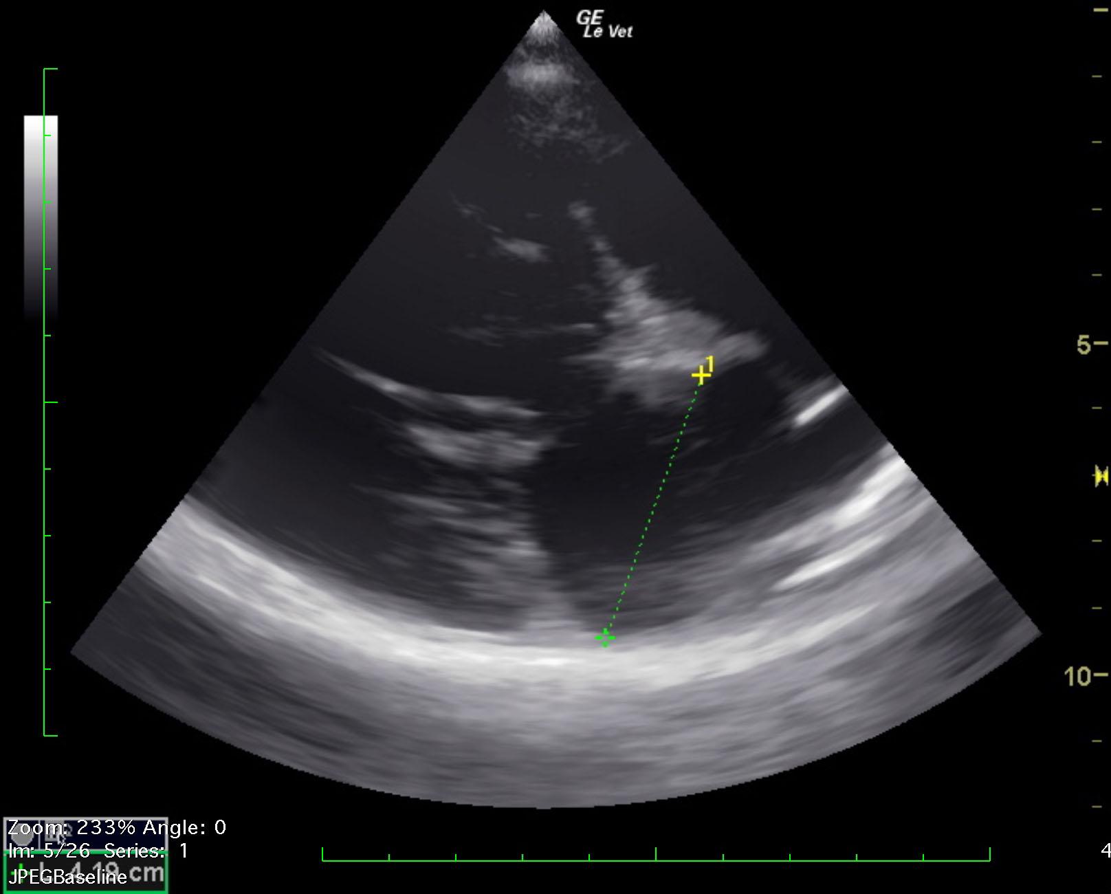
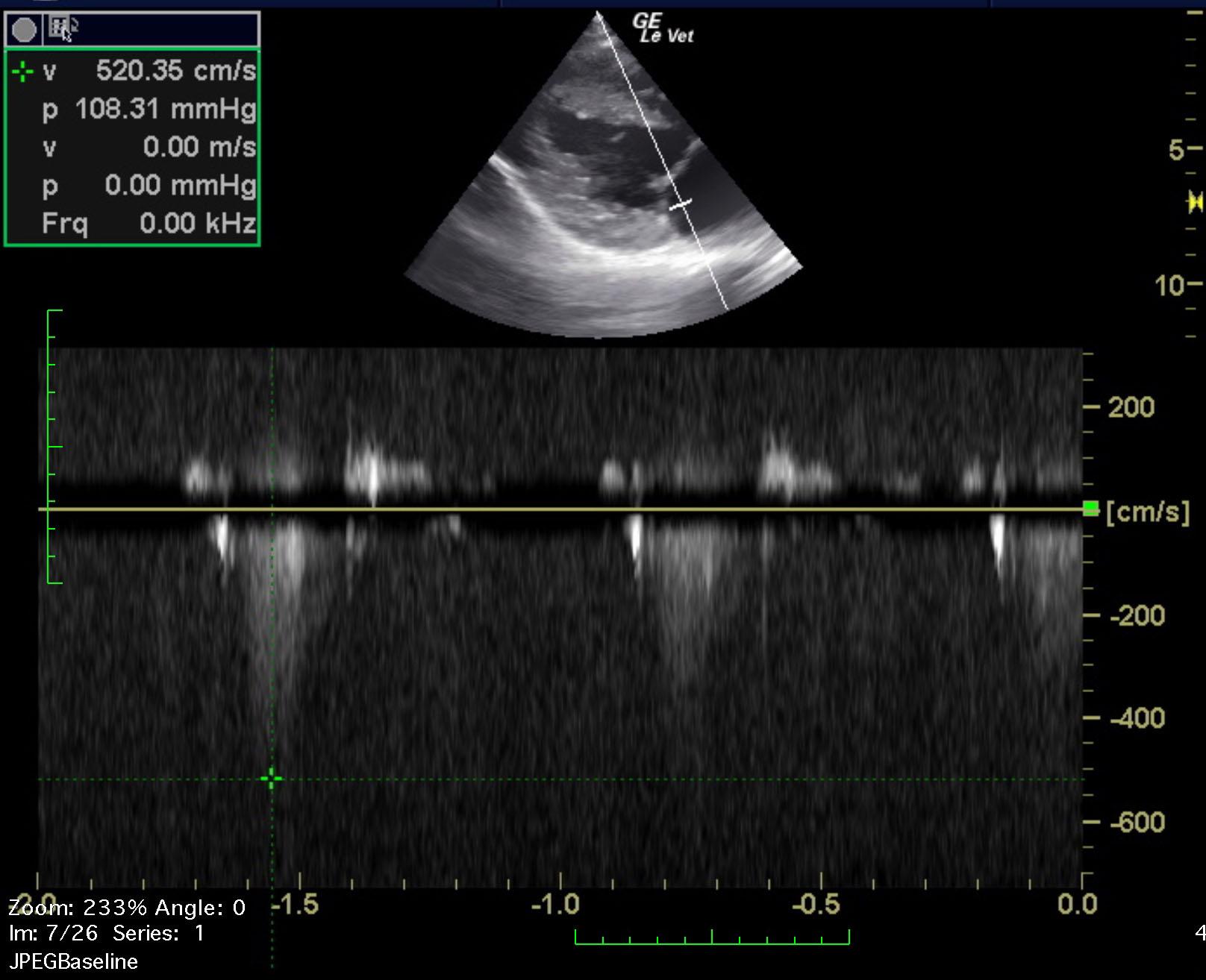
A 3-year-old SF German shepherd was presented for evaluation of a grade IV/VI murmur and normal blood pressure. Survey thoracic radiographs showed thoracic megaesophagus with aspiration pneumonia. On ECG, sinus arrhythmia was evident.


Sonographic Differential Diagnosis

Subaortic Stenosis – severe, concentric hypertrophy present
Aortic Regurgitation – mild

Image Interpretation

The left ventricular chamber measures normal in size. The wall thicknesses are mildly hypertrophied. There is normal systolic function. The left atrium is normal in size (La:Ao 1.30). The visible right heart is normal in size. There is a notable subaortic ridge. Color Doppler identifies an increase in flow and turbulence associated within the suboartic ridge/LVOT. The aortic valve is mildly thickened in some views/grossly normal in other views. The peak velocity of aortic outflow is consistent with severe SAS (LVOT 4.89m/s = 96mmHg). Mild aortic regurgitation is visualized. The mitral valve may be minimally thickened and there is mild MR (peak velocity ~5.20m/s). All other valves appear normal. There is normal, laminar flow across the RVOT/PA (RVOT/PA 1.24m/s). No effusions or masses visualized. IVDd – 1.27cm LVIDd – 4.01cm LVFWd – 1.27cm IVDs – 1.31cm LVIDs – 2.86cm LVFWs – 1.62cm FS – 29%.

DX

Subaortic stenosis, aortic regurgitation

Outcome

None

Clinical Differential Diagnosis

Cardiac – congenital heart disease (aortic stenosis, PDA, pulmonic stenosis, atrial/septal defect), vegetative endocarditis, dilated cardiomyopathy
Congenital/idiopathic megaesophagus

Sampling

None

Video

Patient Information

Gender : Female, Spayed
Species : Canine
Type of Imaging : Ultrasound
Status : For Review

Clinical Signs

  • Heart Murmur

Exam Finding

  • Heart Murmur

Images

img_2269img_2270_la_ao_hbtilly_everett_k9_sas_severe_epsstilly_everett_k9_sas_severe_la_aotilly_everett_k9_sas_severe_lv_mmodetilly_everett_k9_sas_severe_trtilly_sas_severe_ai_sastilly_sas_severe_lembusunnamed_la_maxunnamed

Clinical Signs

  • Heart Murmur