A 14-year-old SF DSH was presented for evaluation of a gallop rhythm. Blood pressure was elevated (168/95). CBC was within normal limits whereas serum biochemistry showed elevated creatinine and ALT activity.
A 14-year-old SF DSH was presented for evaluation of a gallop rhythm. Blood pressure was elevated (168/95). CBC was within normal limits whereas serum biochemistry showed elevated creatinine and ALT activity.
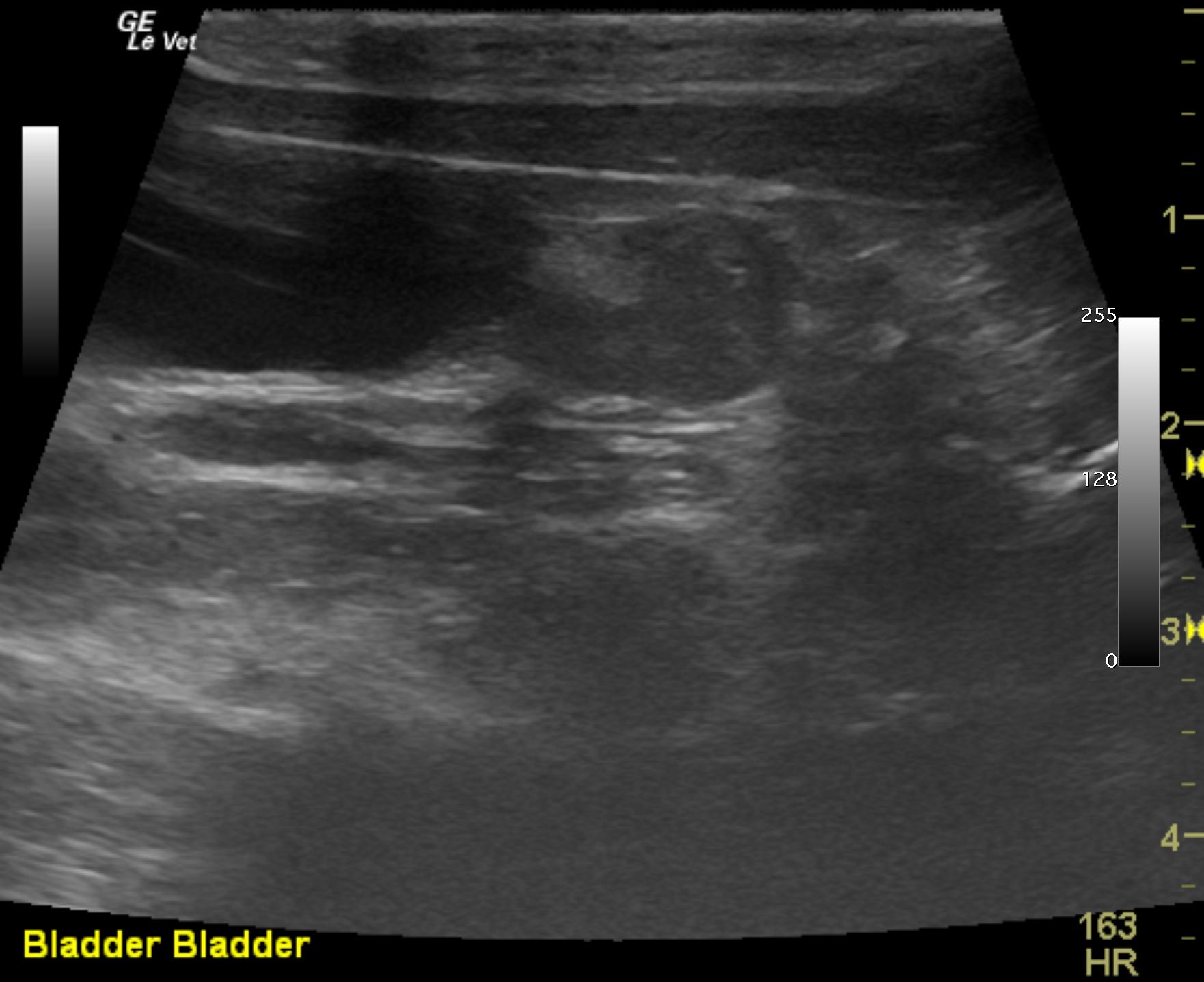
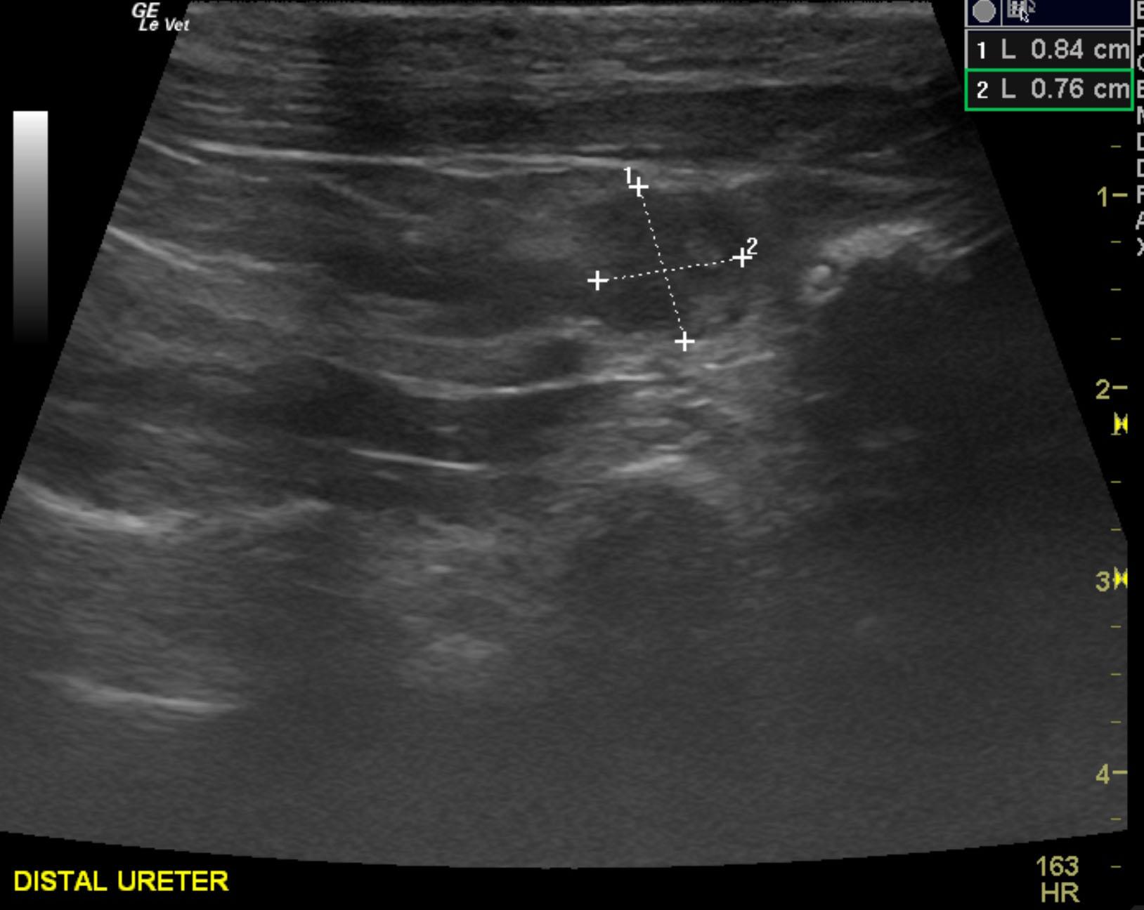
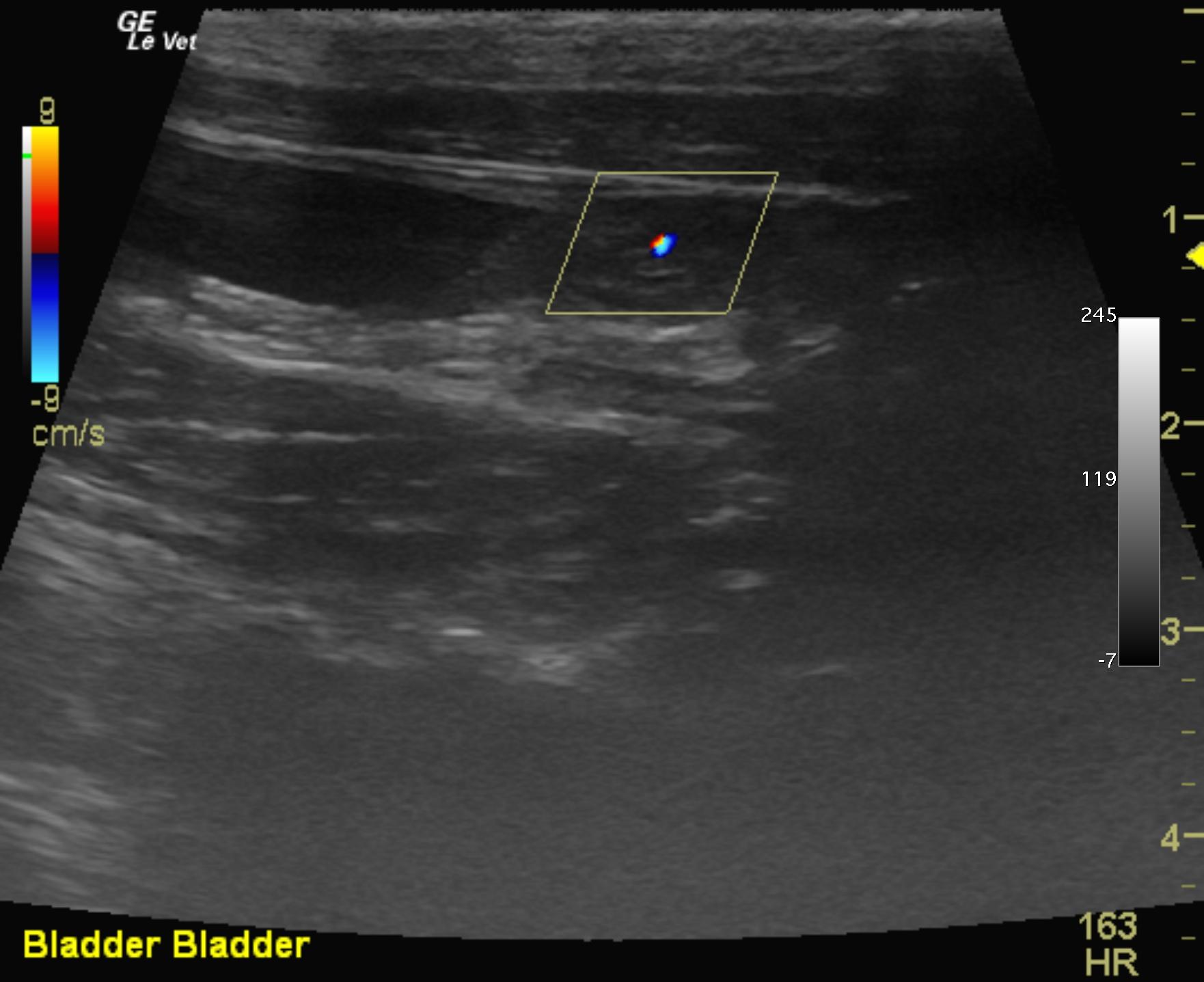
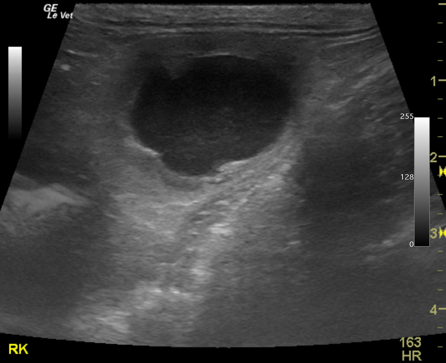
Right ureteral mass. FNA were performed without complication. Suspect transitional cell carcinoma or possible leiomyoma or leiomyosarcoma.
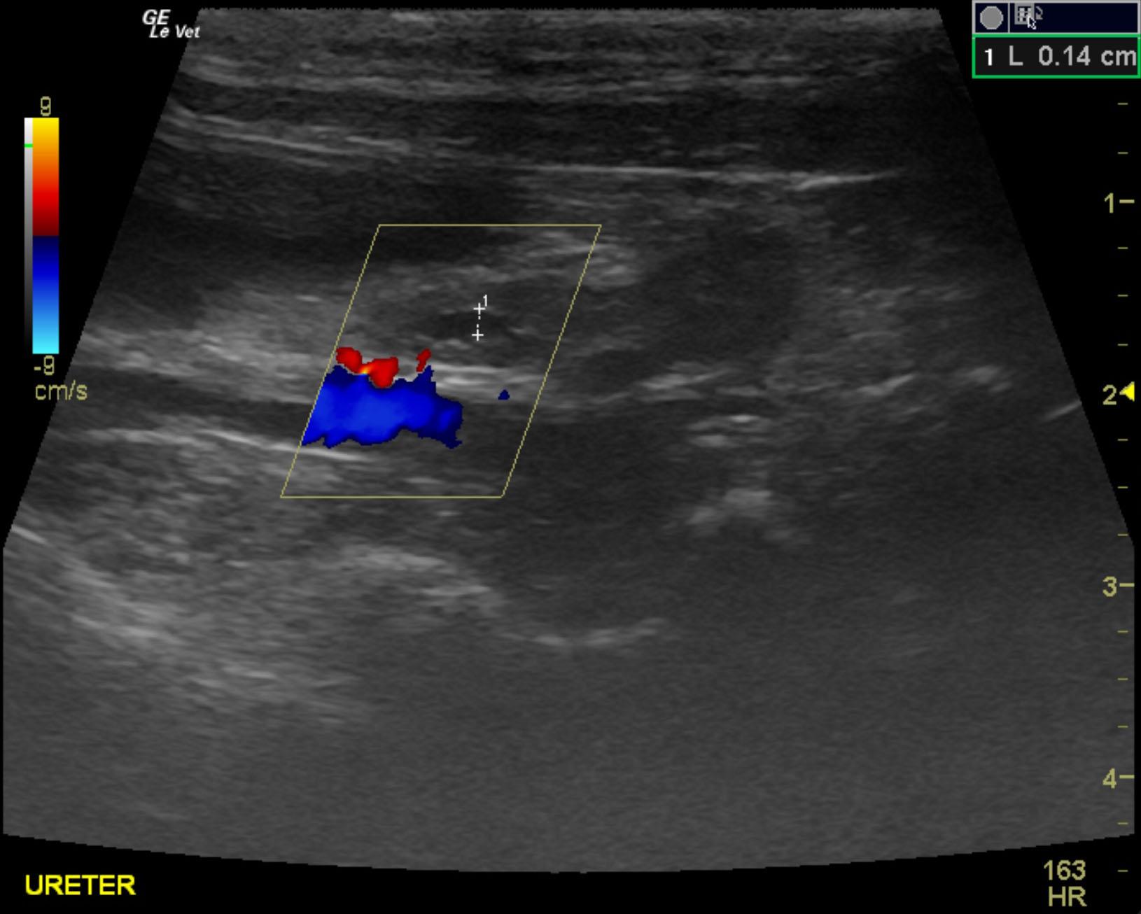
Secondary, moderate, chronic hydronephrosis.
SUB device may be applicable in this case to bypass the ureter and decompress the right kidney. Ultrasound-guided pyelocentesis and decompression of the right kidney can also be considered temporarily to enhance urine flow and relieve pressure upon the right renal nephron. Alternatively surgical intervention could be considered with removal of the distal 4.0 cm of the right ureter and potential reconstruction of the presumed non neoplastic ureter. Otherwise, right nephrectomy could be considered. The left kidney presented mild to moderate degenerative changes. Potential concurrent urinary tract infection, which may be compromising the function. Guarded prognosis.
The right ureter in this patient was thickened at the level of the cystourethral junction as it was obstructed by a mass that measured 0.84 cm at the trigone and infiltrated cranially to approximately 3.0 cm towards the right kidney. Dilation of the right ureter was noted and measured 0.52 cm. The right ureteral mass appeared to enter into the trigone as well. Focal areas of mineralization were noted in the distal ureter, which is suggestive of potential carcinoma. Minor bladder debris was noted. The remaining portion of the bladder was structurally unremarkable.
The right kidney revealed a moderate hydronephrosis echogenic debris, which may be pus. The right kidney measured 3.44 cm. The left kidney presented mild to moderate dystrophic mineralization, slight pyelectasia that measured 0.2 cm. The left kidney also revealed echogenic fat in the renal pelvis and non obstructive calculus. The left kidney measured 2.96 cm, which is subnormal in size.
None
Hypertension – hyperthyroidism, renal disease, adrenal mass, primary
Heart – cardiomyopathy (dilated, hypertrophic, restrictive), myocarditis
FNA of mass yielded…..