A 9-year-old SF DSH with a history of vomiting for years was presented for evaluation of acute deterioration.
A 9-year-old SF DSH with a history of vomiting for years was presented for evaluation of acute deterioration.
A 9-year-old SF DSH with a history of vomiting for years was presented for evaluation of acute deterioration.
A 9-year-old SF DSH with a history of vomiting for years was presented for evaluation of acute deterioration.
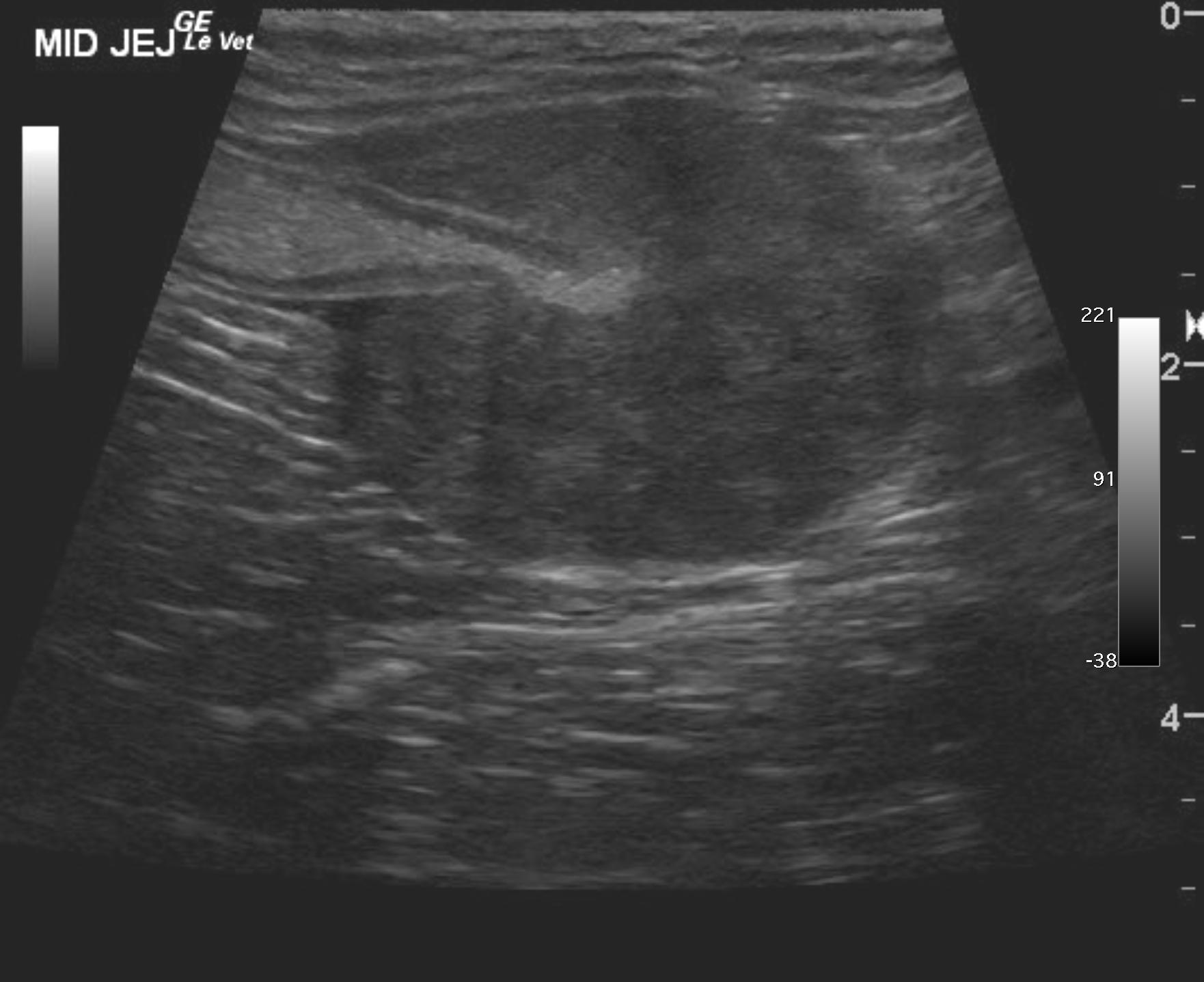
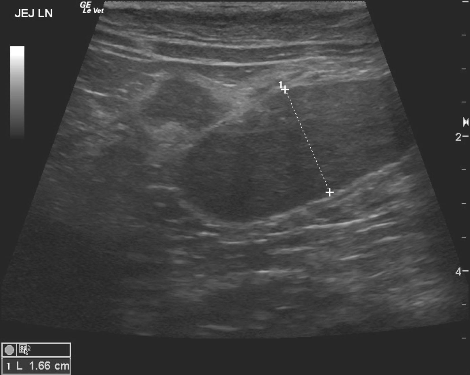
Annular infiltrative jejunal mass with regional lymphadenopathy. Suggestive for focal lymphoma or other round cell neoplasia or adenocarcinoma.
A 2.78 cm wide annular mass was noted in the distal jejunum. Mid-jejunum presented dilation obstructive pattern owing to the mass with retention of chyme. The mass measured approximately 4 cm. Jejunal lymph nodes were enlarged measuring 1.5 cm, hypoechoic. The larger lymph node measured 1.66 cm in width by approximately 3 cm. Some distortion of architecture and pericapsular inflammatory pattern was noted. Given the regional lymphadenopathy, clean resection is not likely possible; however resection and anastamosis would be recommended from the functional standpoint given the obstructive pattern.
None
GIT – neoplasia, IBD, ulceration, foreign body, intussusception, granulomatous enteritis, dietary hypersensitivity, Helicobacter gastritis
Pancreas – pancreatitis, neoplasia
None