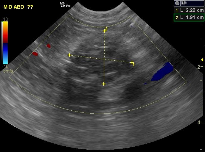
A 3-year-old MN DSH cat was presented for intermittent vomiting. The cat had a previous history of a splenectomy. The cat was BAR on physical examination, however a firm, lobulated, movable mass was palpated in the cranial to mid-abdomen. No other abnormalities were noted. Blood work was unremarkable. Thoracic radiographs showed no abnormalities. Abdominal radiographs showed rounded, amorphous, calcified radiopacities in cranioventral abdomen.
A 3-year-old MN DSH cat was presented for intermittent vomiting. The cat had a previous history of a splenectomy. The cat was BAR on physical examination, however a firm, lobulated, movable mass was palpated in the cranial to mid-abdomen. No other abnormalities were noted. Blood work was unremarkable. Thoracic radiographs showed no abnormalities. Abdominal radiographs showed rounded, amorphous, calcified radiopacities in cranioventral abdomen.
Case Study
Fibrosing enteritis in a 3 year old MN DSH cat
DX
Sonographic Differential Diagnosis
Two separate mid to cranial abdominal structures consistent with foreign material(post surgical, eg sponge) or granulomatous changes with a reactive lymphadenitis. Possible underlying neoplasia.
Image Interpretation
In the area of the mesenteric root, a 2.3cm mixed hypoechoic structure was present with an adjacent hypoechoic structure consistent with lymphadenopathy. The first structure demonstrated a hyperechoic rim consistent with reactive fat. This appears resectable. Cranial to this structure, however a separate shadowing structure was present, the bulk of which appeared associated with the pancreatic limb. Therefore, a partial pancreatic resection may be necessary. These appear resectable and non vascular.
Outcome
The cat was doing well at last communication.
Clinical Differential Diagnosis
GI tract pathology (gastrointestinal foreign body, granuloma, pyogranulomatous lesion (FIP,) abscess, neoplasia – lymphoma, adenocarcinoma, leiomyoma, leiomyosarcoma, mast cell tumor.
Sampling
An exploratory laparotomy was recommended and performed. Biopsies were taken from the mesenteric lymph node, stomach, jejunum, and two firm omental masses. Histopathology revealed fat necrosis with mineralization and fibrosis. No abnormalities were noted on the evaluation of the stomach; however, there was fibrosing enteritis at the level of the jejunum, as well as atypical lymphoid hyperplasia and eosinophilic capsulitis of the mesenteric lymph node.
