03_00530 Trudy B Cirrhosis—-Need captions on cardiac images/video

Case Study

03_00530 Trudy B Cirrhosis—-Need captions on cardiac images/video

A 32 year old, female, African Grey Parrot was presented for a change in attitude and decreased vocalizations after she fell off her perch.  She had an URI one month prior to this presentation and paresis of RT leg for the last month. Findings on chemistry were GGT 35, Albumin 0.9, BUN 2. There was an enlarged heart on radiographs. There was concern for MR and TR and ascites.
 

DX

Cirrhosis

Sonographic Differential Diagnosis

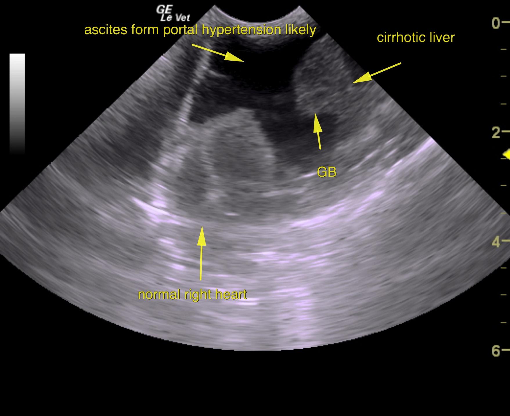
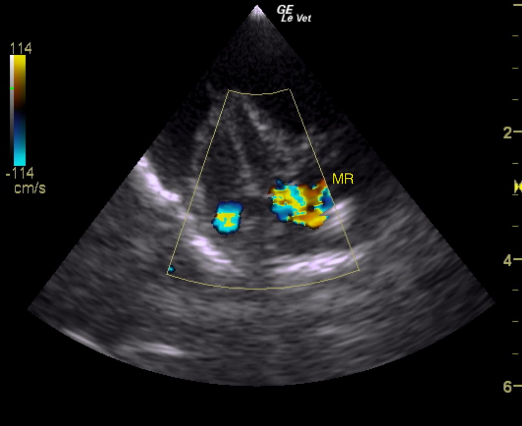
Mild pulmonary hypertension, ascites and cirrhosis pattern. Given the low albumin in this patient, subnormal BUN, elevated liver values and the presence of normal cardiac volume, I feel that the ascites is likely owing from portal hypertension given the severe changes in the liver owing to chronic hepatitis. The mitral and tricuspid insufficiency is likely secondary; however, concurrent pulmonary disease can also be the cause of the mild pulmonary hypertension. Hepatic veins 2 were not dilated nor was the visible vena cava and therefore there is no overt passive congestion pattern that would support ascites deriving from heart failure. Portal hypertension makes the most sense in this patient likely owing to chronic viral disease that may be involving the respiratory tree plus or minus causing the mitral and tricuspid insufficiency.
Prognosis is very guarded.
Medical therapy based on portal hypertension and chronic liver disease would be the preferential approach.

Image Interpretation

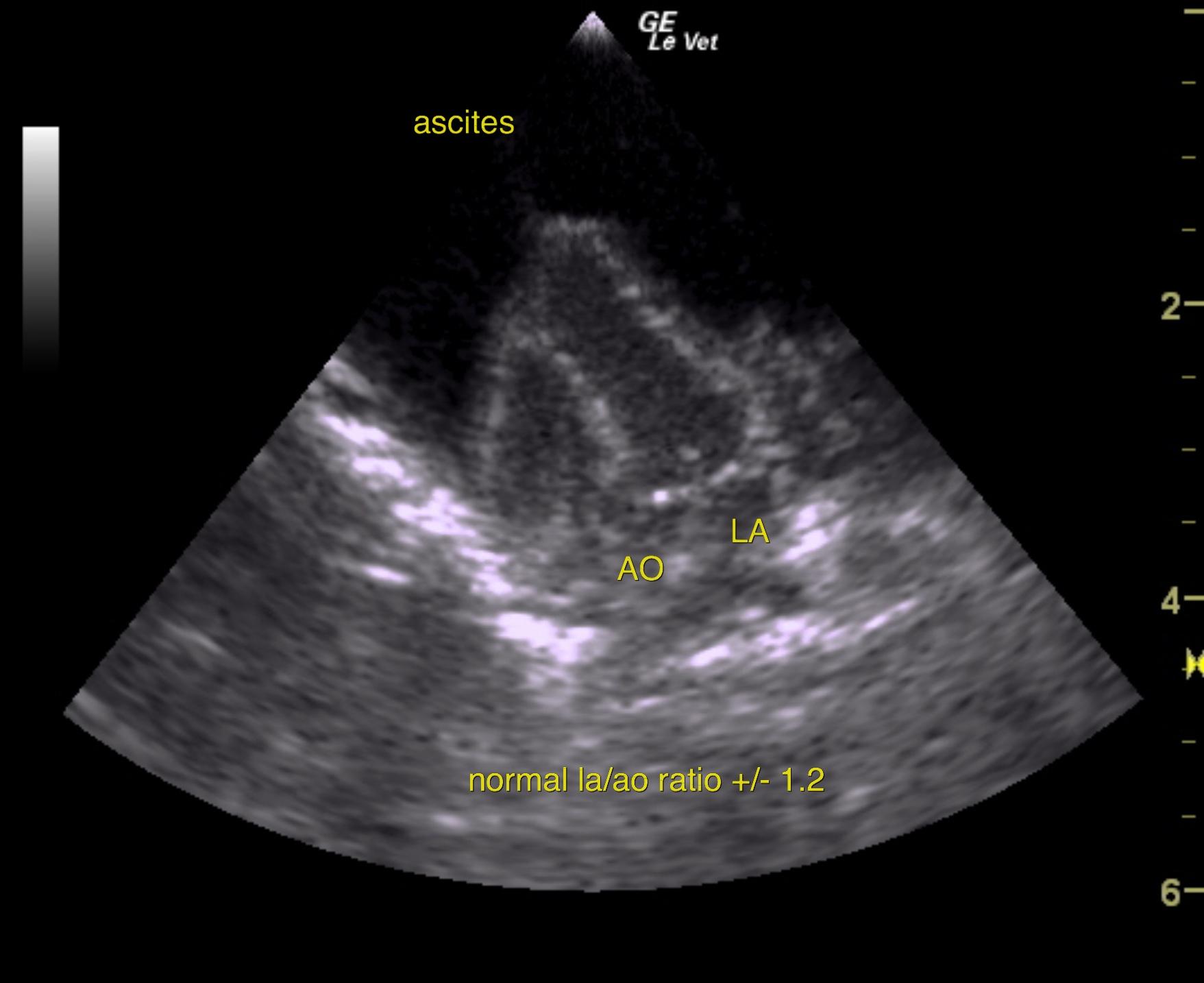
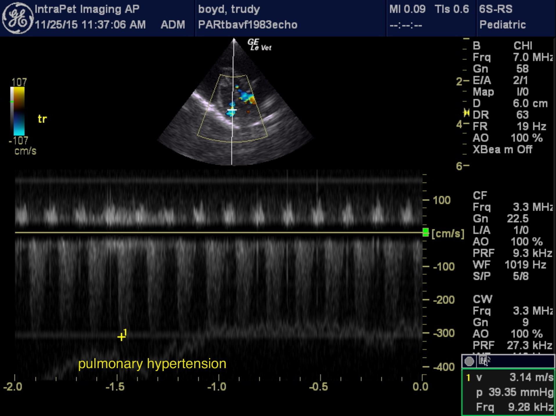
The liver was small, irregular, nodular and coarse in architecture strongly consistent with hepatic cirrhosis. Gallbladder was thickened and collapsed. The cardiac presentation in this patient revealed normal left atrial and right atrial volumes; however, mitral and tricuspid insufficiency was noted. Tricuspid insufficiency was 3.14 m/sec which is consistent with mild pulmonary hypertension. However, the right atrial size and left atrial size were normal. Contractility appeared subjectively adequate. The LA/AO ratio was normal; therefore, no structural evidence of volume overload within the heart itself.
A large amount of ascites was noted around the heart and abdomen.

Outcome

None

Clinical Differential Diagnosis

Cardiac disease, neoplasia, infectious

Sampling

None

Patient Information

Gender : Female, Intact
Species : Exotic
Type of Imaging : Ultrasound
Status : For Review

Clinical Signs

  • "Not Doing Right"
  • Paraparesis

Images

trudy3trudytrudy2trudy5

Blood Chemistry

  • Albumin, Low
  • BUN low
  • GGT High

Clinical Signs

  • "Not Doing Right"
  • Paraparesis
Skip to content