03_00511 Sonni S Passive liver congestion, pleural eff, ascites

Case Study

03_00511 Sonni S Passive liver congestion, pleural eff, ascites

A 10-year-old NM Pomeranian was presented for evaluation of decreased appetite, increased respiratory rate and effort, abdominal distention, and syncopal episodes for the past two weeks. Abnormalities on physical examination were Grade V/VI heart murmur, clear lungs with increased respiratory rate and effort, a palpable fluid wave. Urinalysis showed 3+ proteinuria. Abnormalities on CBC and serum biochemistry were leukocytosis and hypoproteinemia. Survey radiographs showed mild pleural effusion, severe cardiomegaly, and decreased serosal detail within the abdomen.
 

DX

Passive hepatic congestions

Sonographic Differential Diagnosis

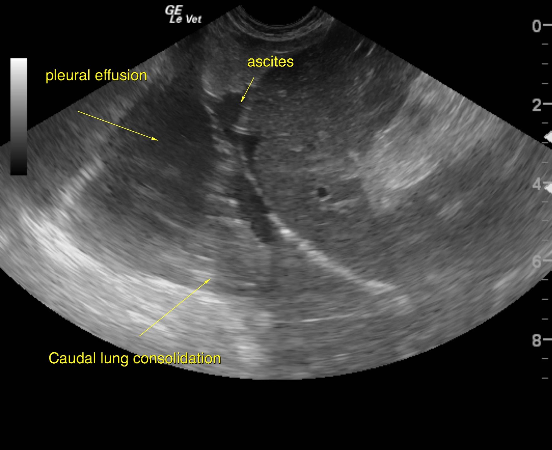
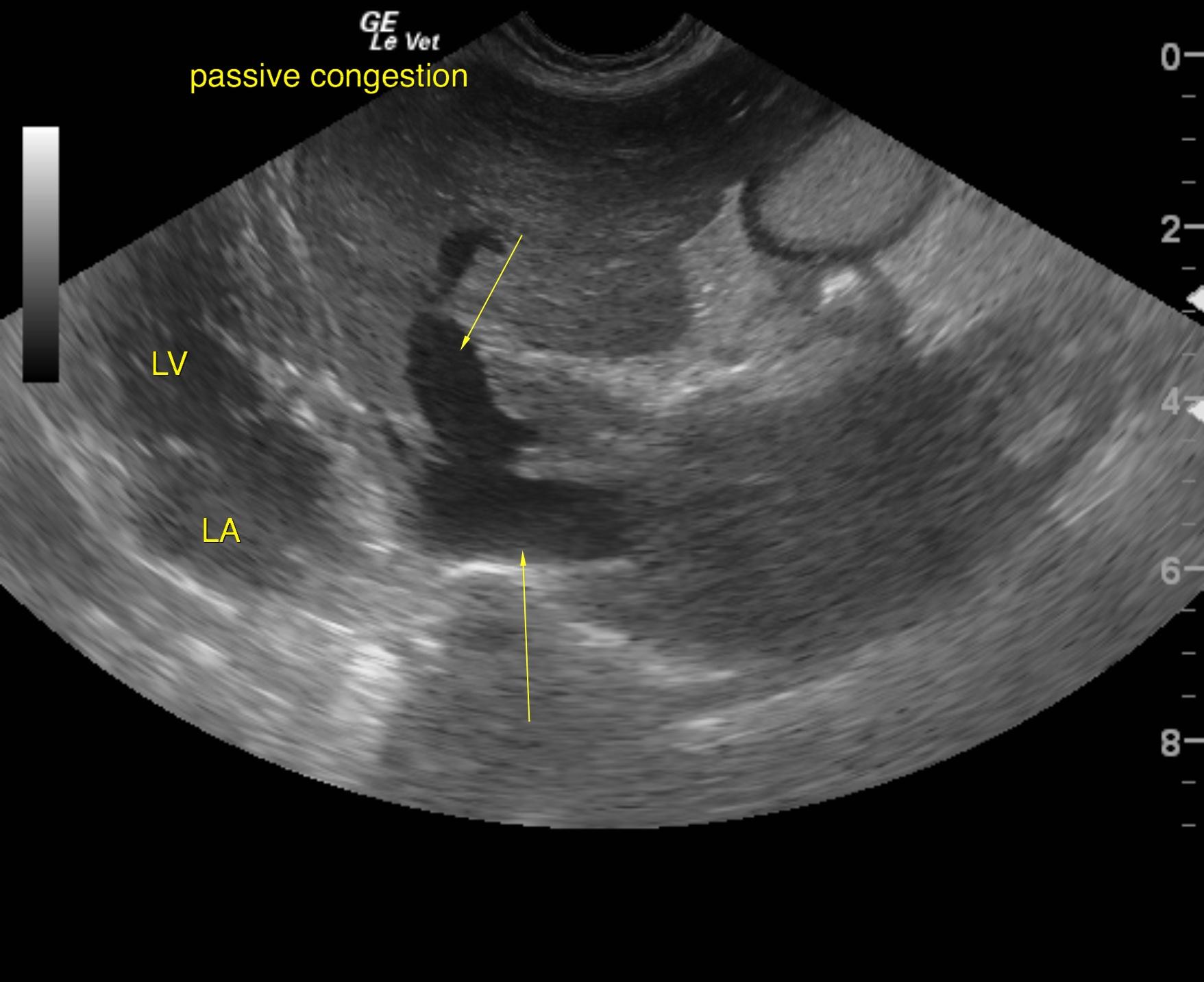
Passive congestion liver pattern with pleural effusion and consolidated lung in the caudal mediastinal region.
I recommend pleurocentesis in this patient to assess for carcinoma or similar neoplastic cells that may be enveloping the thoracic duct, vena cava and caudal lung field. The abdominal pattern is most consistent with passive congestion and secondary ascites. An echocardiogram is warranted. However, typically right-sided heart failure does not regularly cause pleural effusion. Intercostal approach to the sonogram is recommended in the thorax to assess for lung consolidation and potential aspiration. However, pleurocentesis with cytospin and immediate slide preparation may conserve cells for adequate cellular diagnosis.

Image Interpretation

The liver was uniformly swollen. The hepatic veins were dilated in this patient as was the vena cava. This is consistent with passive congestion pattern.
A moderate amount of ascites was noted. Echogenic fat was noted throughout the abdomen, yet this is artifactual owing to the ascites.
Pleural effusion was also noted with consolidated lung.

Outcome

None

Clinical Differential Diagnosis

Heart – valvular endocardiosis, vegetative endocardiosis, myocardial failure
Liver – cirrhosis, neoplasia
Glomerulonephritis

Sampling

None

Patient Information

Gender : Male, Neutered
Species : Canine
Type of Imaging : Ultrasound
Status : Complete

Clinical Signs

  • Abdominal Distension
  • Anorexia
  • Syncopal episodes
  • Tachypnea

Exam Finding

  • Free fluid
  • Heart Murmur

Images

son1son2

Blood Chemistry

  • Total Protein, Low

CBC

  • WBC, High

Clinical Signs

  • Abdominal Distension
  • Anorexia
  • Syncopal episodes
  • Tachypnea

Urinalysi

  • Protein Present
Skip to content