A 13-year-old MN Shetland Sheepdog with lengthy history of gastrointestinal issues, cholecystectomy 1 year prior, and elevated liver enzymes, was presented for limping. On physical examination the dog had right fore lameness and an enlarged left elbow was evident. The only significant abnormality on blood chemistry was severely elevated ALP activity (7125 U/L). Elbow radiographs were diagnostic for severe osteoarthritis. An adrenal panel from the University of Tennessee indicated the presence of moderately increased adrenal activity.
A 13-year-old MN Shetland Sheepdog with lengthy history of gastrointestinal issues, cholecystectomy 1 year prior, and elevated liver enzymes, was presented for limping. On physical examination the dog had right fore lameness and an enlarged left elbow was evident. The only significant abnormality on blood chemistry was severely elevated ALP activity (7125 U/L). Elbow radiographs were diagnostic for severe osteoarthritis. An adrenal panel from the University of Tennessee indicated the presence of moderately increased adrenal activity.
Case Study
03_00203 Mozart D Hepatic mass: carcinoma, pseudomonas infection
DX
Sonographic Differential Diagnosis
Periportal mass. Nodular hyperplasia of the liver. Low grade pancreatic and gastric inflammation.
Image Interpretation
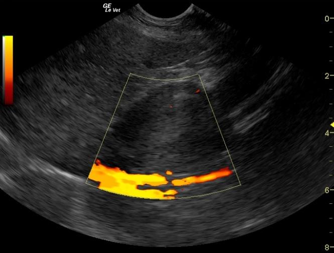
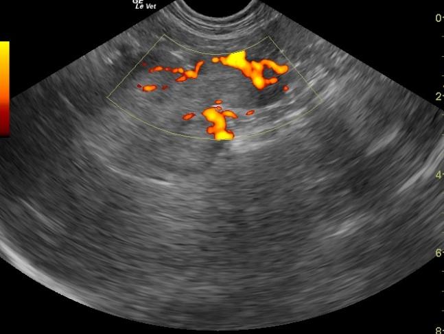
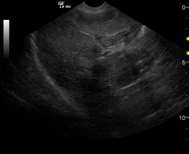
The liver in this patient presented swollen contour with micro and macronodular changes. A large periportal mass was noted. This deviated the common bile duct and portal vein. This was significantly vascular. This mass either derived from the periportal lymph nodes or possibly the pancreatic tissue as it appears contiguous with pancreatic tissue. This mass was found between and dorsal to the portal vein. It also deviated the vena cava dorsally. The stomach presented prominent mucosa. This is consistent with low grade gastritis. The pancreas presented mixed hypoechoic changes in the right pancreatic limb. This is consistent with low grade pancreatitis or possible nodular hyperplasia. The remainder of the pancreas presented minor age related changes.
Clinical Differential Diagnosis
Elbow DJD Elevated ALP – Cushing’s disease, cholestasis, pancreatic disease, bone metabolism
Sampling
US-guided FNA with a 25 gauge needle performed of the abdominal mass near the caudal border of the liver near the pancreas revealed vacuolar hepatopathy with atypical epithelial proliferation; suggestive of carcinoma, lymphocytic and neutrophilic inflammation; blood and lipid. Culture of the mass revealed pseudomonas.
Video
Patient Information
Clinical Signs
- Lameness
History
- Cholecystectomy
- Elevated Liver Enzymes
- GI Issues
Images
Blood Chemistry
- Alkaline Phosphatase (SAP), High
Clinical Signs
- Lameness