A 4-year-old F Shih Tzu was presented for loss of bladder control, lethargy, and PU/PD for one month. Abnormalities on physical examination were pyrexia (104° F), depressed, and a painful abdomen on palpation. CBC and blood chemistry showed leukocytosis, low MCV, neutrophilia, hypoglycemia, hypocalcemia, and hypomagnesemia. On survey abdominal radiographs renomegaly, urinary calculi, and decreased abdominal detail with some possible liver involvement was evident. The patient was treated with I.V. fluids pending further diagnostics.
A 4-year-old F Shih Tzu was presented for loss of bladder control, lethargy, and PU/PD for one month. Abnormalities on physical examination were pyrexia (104° F), depressed, and a painful abdomen on palpation. CBC and blood chemistry showed leukocytosis, low MCV, neutrophilia, hypoglycemia, hypocalcemia, and hypomagnesemia. On survey abdominal radiographs renomegaly, urinary calculi, and decreased abdominal detail with some possible liver involvement was evident. The patient was treated with I.V. fluids pending further diagnostics.
Case Study
Splenoazygos shunt in a 4 year old F Shih Tzu dog
DX
Sonographic Differential Diagnosis
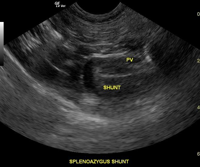
Large splenoazygos shunt which measured 1.2cm at maximum width. Aggressive acute nephritis of the right kidney likely due to passage of large nephrolith. Multiple large urinary bladder calculi. Stable left kidney calculi.
Image Interpretation
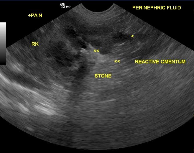
The urinary bladder presented multiple strongly shadowing calculi. The bladder wall was within normal limits. The urethra and ureters were not obstructed at the level of the urinary bladder. The left kidney was mildly swollen in contour measuring 5.7cm and the right kidney measured 6.4cm. The left kidney did not appear inflamed, however medullary calculi were present and numerous. No hydronephrosis was noted. The right kidney presented multiple medullary calculi, swollen ill defined cortices and poor corticomedullary definition. Some echogenic perinephric fluid was noted that continued along the fascial plane highly suggestive for either hemorrhage or urine or a combination. This is suggestive for aggressive nephritis likely due to passage of calculi from the kidney to the urinary bladder. No hydroureter was noted at this time. The liver was subnormal in size at 2.4cm in maximum width in short axis. A 1.2cm splenoazygos shunt was present. The pre-shunt portal vein measured 0.45cm and post shunt portal vein measured 0.25cm. The caudal vena cava and aorta in the same position measured 0.5cm each highly suggestive for splenoazygos shunt.
Outcome
The patient was humanely euthanized.
Clinical Differential Diagnosis
Multi-organ pathology – Renal infection/pyelonephritis/renal failure with UTI, liver failure, renal/bladder calculi, peritonitis – bacterial/urine, septicemia
Video
Patient Information
Clinical Signs
- Incontinence
- Lethargy
- PU-PD
Exam Finding
- Abdominal Pain
- Depression
- Fever
Blood Chemistry
- Calcium, Low
- Cholesterol, High
- Glucose, Low
- Magnesium, Low
CBC
- MCV, High
- Neutrophils, High
- WBC, High
Clinical Signs
- Incontinence
- Lethargy
- PU-PD
Urinalysi
- Albumin Present

