A 9-year-old MN Rottweiler dog presented for progressively worsening anorexia and mild weight loss. Preliminary diagnostic testing revealed moderate anemia with slight regeneration, anisocytosis, 24 percent band neutrophils, Dohle bodies, normal total WBC count, and mildly elevated SAP and mildly elevated AST.
A 9-year-old MN Rottweiler dog presented for progressively worsening anorexia and mild weight loss. Preliminary diagnostic testing revealed moderate anemia with slight regeneration, anisocytosis, 24 percent band neutrophils, Dohle bodies, normal total WBC count, and mildly elevated SAP and mildly elevated AST.
Case Study
03_00039 Chipper K Malignant histiocytosis, vascular and biliary ectasia
DX
Malignant histiocytosis with vascular and biliary ectasia
Sonographic Differential Diagnosis
Primary consideration is given to neoplasia, most likely of round cell origin. An acute inflammatory hepatopathy cannot be ruled out; however, it is considered less likely given the portal vascular changes.
Image Interpretation
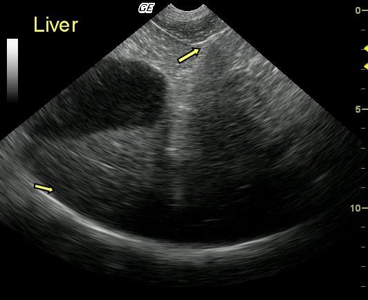
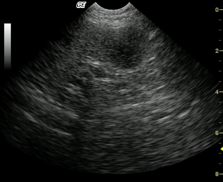
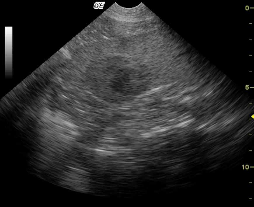
Transverse view of the left liver and gallbladder reveals moderate hepatic enlargement with diffusely and moderately hypoechoic parenchyma and evidence of reduced vascular volume and sound attenuation (thin arrow). Diffuse intraparenchymal portal vascular displacement and, in the near field, a swollen serosal margin are present (wide arrow) adjacent to the falciform ligament.
Outcome
The patient was treated with immune suppressive cortisone because the owner declined chemotherapy. The patient was humanely euthanized after severe weight loss and clinical decline 1 month post-diagnosis.
Comments
Similar findings were derived from the splenic biopsy, which further supported the pathologist’s ultimate diagnosis of systemic malignant histiocytosis.
Clinical Differential Diagnosis
Systemic infection, neoplasia, hepatitis, Addison`s disease, GI blood loss.
Sampling
16-gauge US-guided biopsy revealed malignant histiocytosis with vascular and biliary ectasia.


