Renal hemangiosarcoma in a 5-year-old intact male German Shepherd: Our Case Of the Month March 2015

Case Of the Month

Renal hemangiosarcoma in a 5-year-old intact male German Shepherd: Our Case Of the Month March 2015

A 5-year-old male German shepherd was presented for evaluation of progressive pain, lethargy, anorexia, and hunched back following an episode of abdominal trauma – tried to jump over a large hole and hit his abdomen on the edge of the hole.

A 5-year-old male German shepherd was presented for evaluation of progressive pain, lethargy, anorexia, and hunched back following an episode of abdominal trauma – tried to jump over a large hole and hit his abdomen on the edge of the hole.
DX

Renal hemangiosarcoma and subcapsular hematoma

Outcome

Nephrectomy revealed renal mass with hemangiosarcoma confirmed upon histopathological review.

Comments

Hemangiosarcoma is a rapidly expanding neoplastic process that can often present with a history of suspected “trauma.” Given that both traumatic lesions and HSA can cause hematoma formation HSA should be considered especially in predisposed breeds. Traumatic clot should begin to resolve progressively but logically HSA will progress and expand as it did in this case.


Sampling

Sampling: Surgical biopsy revealed renal hemangiosarcoma and subcapsular hematoma.

Sonographic Differential Diagnosis

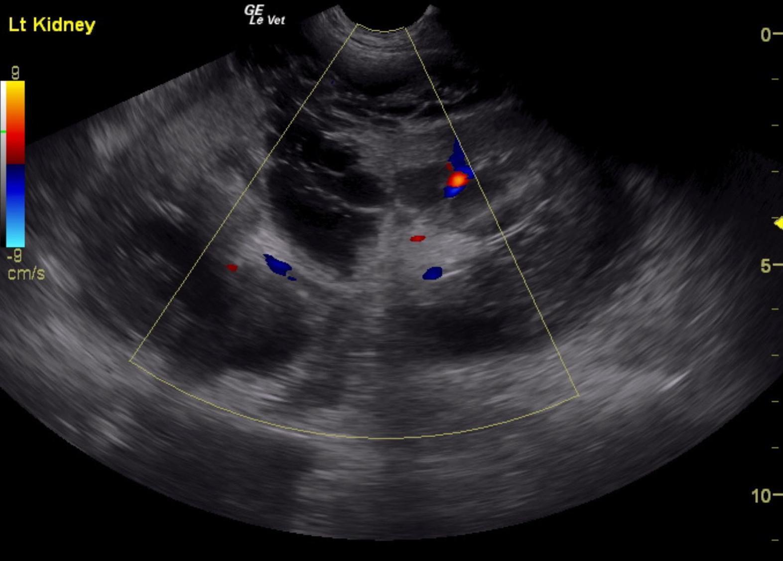
Sonographic Differential Diagnosis: Renal hemorrhage with pericapsular and retroperitoneal fluid and tissue proliferation. Potential clot versus neoplasia and secondary hemorrhage. FNA of the hypoechoic tissue is recommended. Guarded prognosis. Recheck sonogram is recommended in 48-72 hours to assess for any progression or resolution. Neoplastic infiltrates and blood clots can look similar on ultrasound. Doppler assessment could not differentiate one or the other hence the necessity for aspirates.

Image Interpretation

Sonographic Interpretation:. The left kidney in this patient presented a subcapsular blood clot and hemorrhage escape or possible tumor related escape through the dorsal cortex. This appears to extend outside the capsule. Tissue thickening was noted and extended caudally between the vena cava and aorta. This may represent hemangiosarcoma or other neoplastic event with capsular escape. FNA of the hypoechoic tissue and perirenal space and sublumbar space is recommended to assess for a potential neoplastic event and secondary hemorrhage. Otherwise, exploratory surgery is recommended. However, the patterns extending caudally in the sublumbar and retroperitoneal space will not likely allow for clean resection. The left kidney was severely enlarged at 10.6 cm. Infiltrative process is suspected with potential for a blood clot.

Patient Information

Patient Name : Rocky Galloway
Gender : Male, Intact
Species : Canine
Status : Complete

Clinical Signs

  • "Not Doing Right"
  • Abdominal Pain
  • Anorexia
  • Lethargy

Exam Finding

  • Abdominal Pain
  • Lethargy

Images

rocky_galloway_lk_hsa_1_post_copyrocky_galloway_lk_hsa_post_copyrocky_galloway_lk_hsa_original_12_5_prior_to_med_tx_2_copyrocky_galloway_rk_normal_copy

Clinical Signs

  • "Not Doing Right"
  • Abdominal Pain
  • Anorexia
  • Lethargy
Skip to content