Skip to content
Customize Consent Preferences

We use cookies to help you navigate efficiently and perform certain functions. You will find detailed information about all cookies under each consent category below.

The cookies that are categorized as "Necessary" are stored on your browser as they are essential for enabling the basic functionalities of the site. ... 

Always Active

Necessary cookies are required to enable the basic features of this site, such as providing secure log-in or adjusting your consent preferences. These cookies do not store any personally identifiable data.

No cookies to display.

Functional cookies help perform certain functionalities like sharing the content of the website on social media platforms, collecting feedback, and other third-party features.

No cookies to display.

Analytical cookies are used to understand how visitors interact with the website. These cookies help provide information on metrics such as the number of visitors, bounce rate, traffic source, etc.

No cookies to display.

Performance cookies are used to understand and analyze the key performance indexes of the website which helps in delivering a better user experience for the visitors.

No cookies to display.

Advertisement cookies are used to provide visitors with customized advertisements based on the pages you visited previously and to analyze the effectiveness of the ad campaigns.

No cookies to display.

Intraoperative Ultrasound

Case Of the Month

Intraoperative Ultrasound

We developed this technique to be used on any abdominal organ but is especially effective in case of infiltrative, focal and multifocal GI lesions. The problem is that the surgeon cannot often see what the clinical sonographer is observing from a transabdominal sonographic perspective. If the organ serosa is not visibly affected, the surgeon will simply perform a “shopping spree” of intestinal biopsies as opposed to a precise sampling procedure of the most representative lesion that we observe sonographically.

We developed this technique to be used on any abdominal organ but is especially effective in case of infiltrative, focal and multifocal GI lesions. The problem is that the surgeon cannot often see what the clinical sonographer is observing from a transabdominal sonographic perspective. If the organ serosa is not visibly affected, the surgeon will simply perform a “shopping spree” of intestinal biopsies as opposed to a precise sampling procedure of the most representative lesion that we observe sonographically. Hence we may identify and resect the most representative mural lesions with this method.

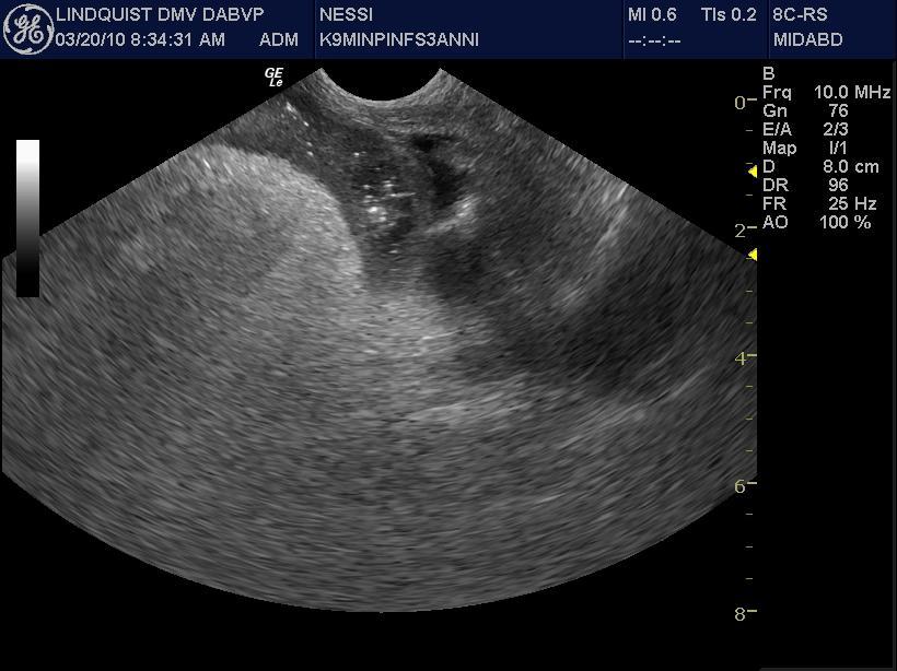
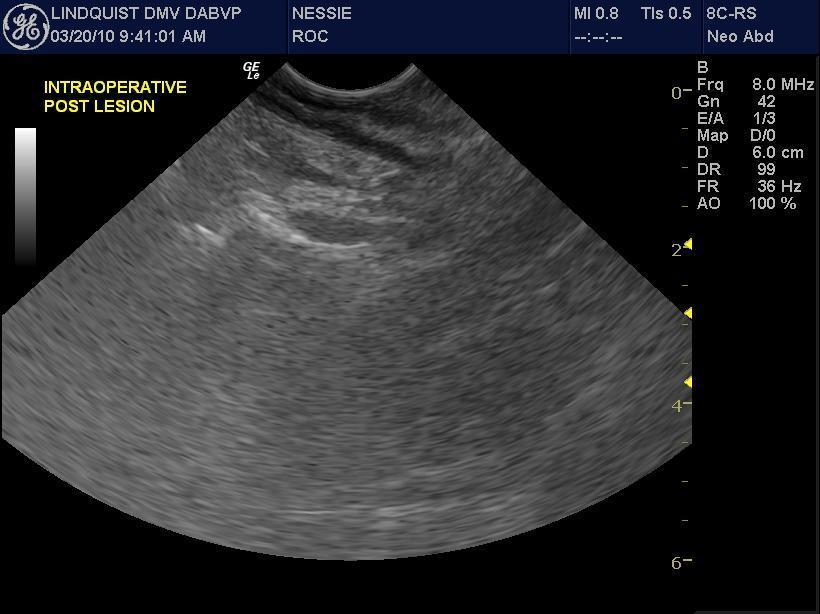
Procedure: Acoustic gel is placed into a double surgical glove to keep the outside exposed glove sterile. Cold sterilize the ultrasound probe with alcohol before putting it in the glove. Pull the glove tight on top the probe to ensure adequate probe/gel/glove coupling occurs to avoid any air entrapment. Have the surgeon exteriorize the bowel or expose the target organ to be sampled. A technician pours saline on the bowel (or other organ) as a coupling agent. Scan the organ to define the most representative region of the mural pathology that was observed transabdominally. Then define the best healthy tissue where the infiltrative pattern or pathology subsides and resect the lesion at this identified point of healthy tissue proximal and distal to the affected region. This procedure should take the sonographer 10 minutes or so and the surgeon may do the rest.

Comments

This procedure ensures as complete a resection of unhealthy bowel as possible and also ensures that healthy bowel is utilized for the anastamosis. Postoperative dehiscence may be avoided by this intraoperative intervention. More on this technique may be seen in our abstract from ECVIM 2009 (Intraoperative Ultrasound for Precise Biopsy and Resection of Transabdominally Detected Intestinal Lesions in 3 cats. Lindquist, Casey, Frank)

Patient Information

Images

res_int_intraoperative_ultrasound_01res_int_intraoperative_ultrasound_04