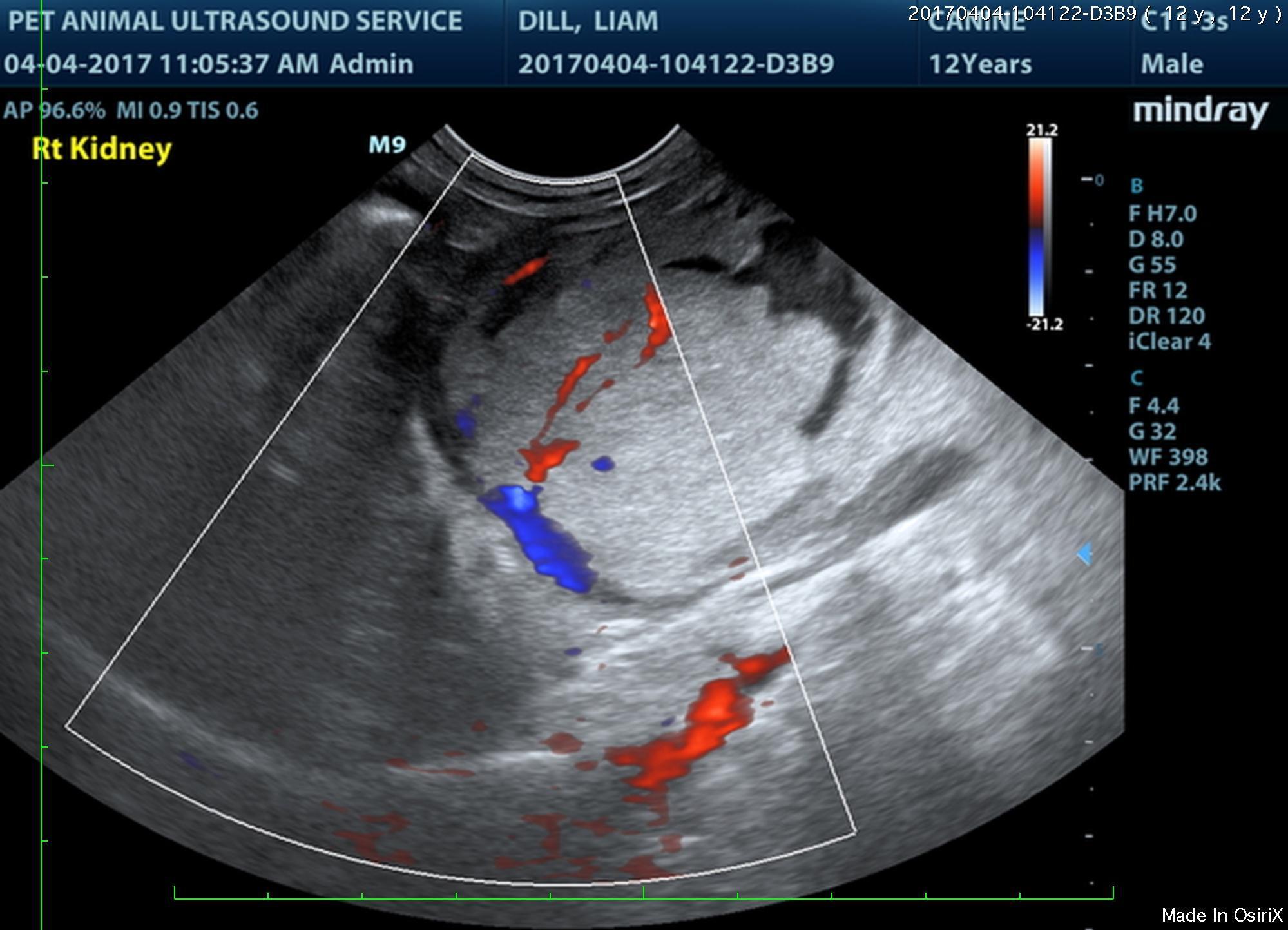
A 12-year-old NM Jack Russell Terrier was presented for evaluation of hematuria with one episode of stranguria. Urinalysis showed specific gravity of 1.032, 3+ blood, and 4+ protein. CBC and serum biochemistry was within reference range.
A 12-year-old NM Jack Russell Terrier was presented for evaluation of hematuria with one episode of stranguria. Urinalysis showed specific gravity of 1.032, 3+ blood, and 4+ protein. CBC and serum biochemistry was within reference range.