CT – Primary nasal neoplasia in a 10 year old FS Chow Chow

Case Study

CT – Primary nasal neoplasia in a 10 year old FS Chow Chow

This 10 year old FS Chow Chow has chronic rhinitis, episodic right-sided epistaxis

This 10 year old FS Chow Chow has chronic rhinitis, episodic right-sided epistaxis

Image Interpretation

CT of the skull-

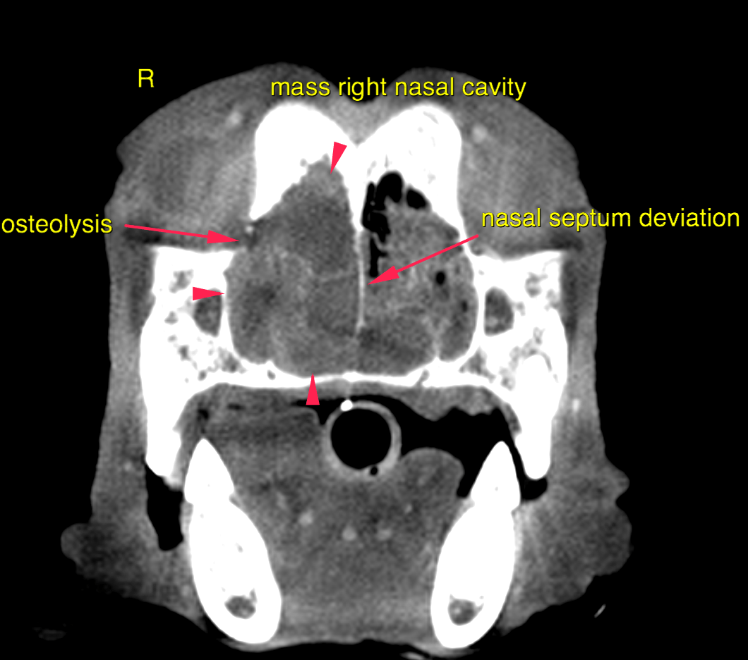
The right nasal cavity and frontal sinus are filled by a mildly heterogeneous soft tissue attenuating and irregularly contrast enhancing mass. Multifocal amorphous mineral attenuating areas are noted within the mass. The mass perforates into the left nasal cavity and there is complete obstruction of the ventral and middle nasal meatus on the left side. The nasal septum presents lysis and moderate deviation to the left side. Complete destruction of the conchal and turbinate structures is seen in the right nasal cavity associated with the mass. Partial destruction is noted in the left nasal cavity. The pars orbitalis of the frontal bone presents regional permeative osteolysis.  One of the right submandibular lymph nodes on the right side is markedly enlarged and rounded.

DX

The findings are consistent with a primary nasal neoplasia.

Outcome

Most likely differentials
include adenocarcinoma, lymphoma, squamous cell carcinoma, other.
Suspicion for metastatic spread to one of the right submandibular lymph nodes. Consider follow-up rhinoscopy with repetition of the biopsy as the preferred diagnostic
method. Depending on the results of histopathological results radiation-therapy would
be treatment option of choice. FNA samples of the enlarged submandibular lymph
node are essential.
Patient Information

Patient Name : Cinnamon Sherrer/MPI
Gender : Female, Spayed
Species : Canine
Type of Imaging : Ultrasound
Status : Complete
Liz Wuz Here : Yes
Code : 16_00139

Clinical Signs

  • Nasal discharge bloody

Images

bildschirmfoto_2016-09-02_um_11

Clinical Signs

  • Nasal discharge bloody