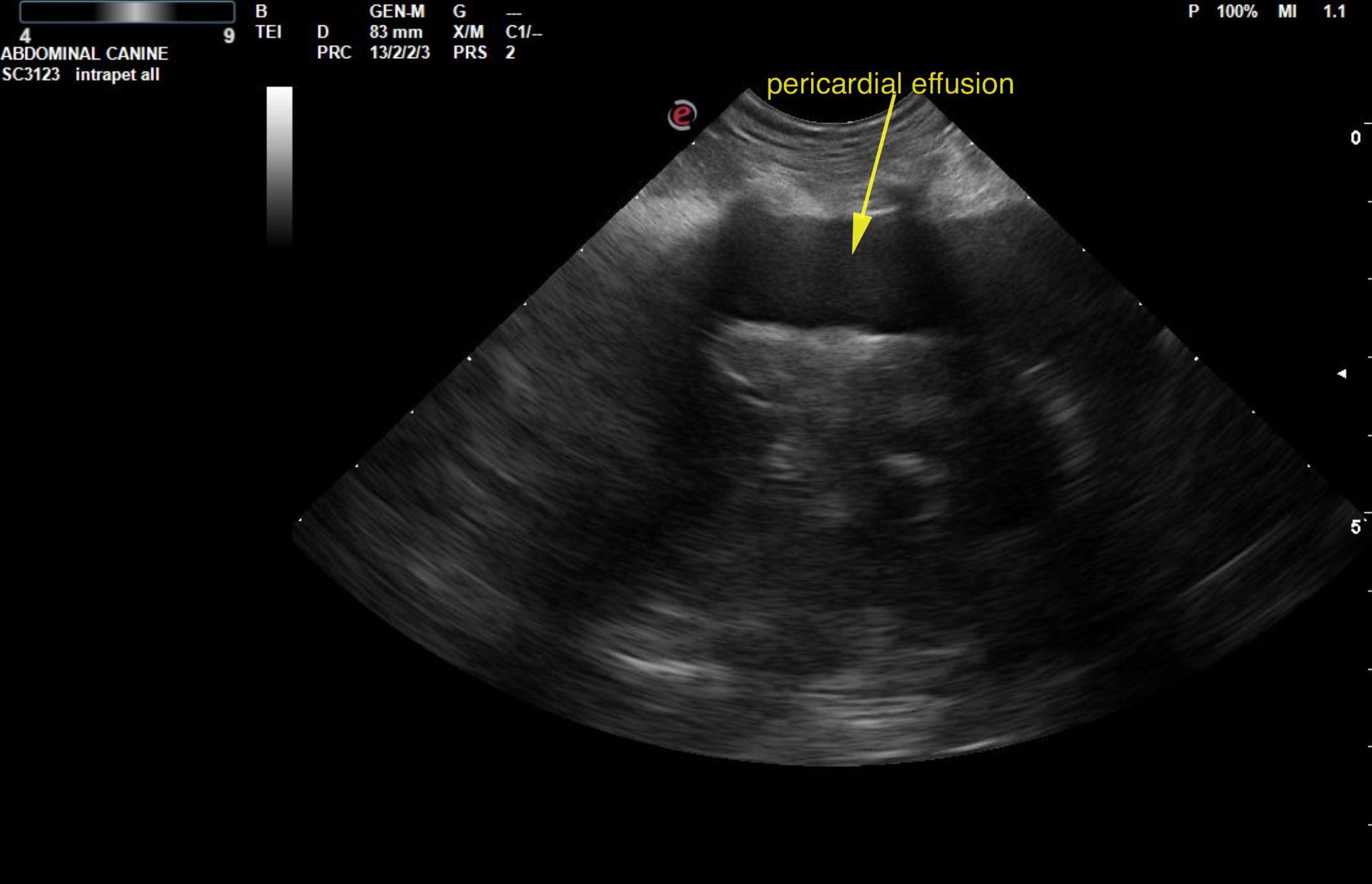
This 12 year old MN Dachshund dog presented with vomiting, inappetance, distended abdomen.
Rads showed cardiac enlargement, pulmonary edema
CBC/Chem: ALT 39, ALKP 188, WBC 23,000
This 12 year old MN Dachshund dog presented with vomiting, inappetance, distended abdomen.
Rads showed cardiac enlargement, pulmonary edema
CBC/Chem: ALT 39, ALKP 188, WBC 23,000