This 14 year old Siberian Husky dog has a history of grand mal seizures of one yar duration. Presented with focal seizures. Large growth noted on the abdomen
Physical exam: Presented laterally recumbant. Acute ataxia with nystagmus and head tilt
CBC/CHem: HCT 32%, Ca low
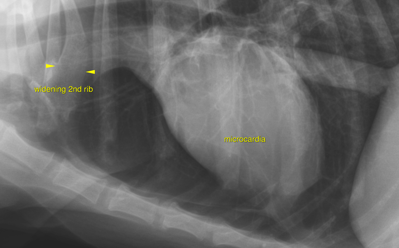
Looking for mets
This 14 year old Siberian Husky dog has a history of grand mal seizures of one yar duration. Presented with focal seizures. Large growth noted on the abdomen
Physical exam: Presented laterally recumbant. Acute ataxia with nystagmus and head tilt
CBC/CHem: HCT 32%, Ca low
Looking for mets