ORTHO – Chronic bilateral tendovaginitis of the biceps in a 6 year old MN Australian Cattle Dog

Case Study

ORTHO – Chronic bilateral tendovaginitis of the biceps in a 6 year old MN Australian Cattle Dog

This 6 year old MN Australian Cattle Dog presented with mild left forelimb lameness of 3-4 months duration

Physical exam: decreased carpal flexion with mild effusion, mild discomfort noted on the medial aspect of the elbow but normal ROM. Spasm of left biceps tendon stretch.
This 6 year old MN Australian Cattle Dog presented with mild left forelimb lameness of 3-4 months duration

Physical exam: decreased carpal flexion with mild effusion, mild discomfort noted on the medial aspect of the elbow but normal ROM. Spasm of left biceps tendon stretch.
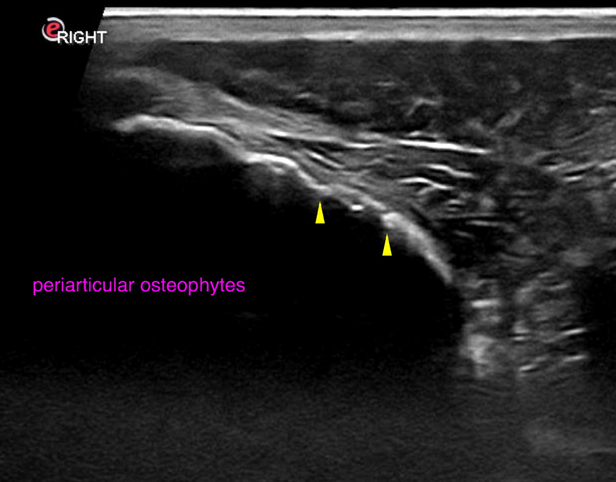
Image Interpretation

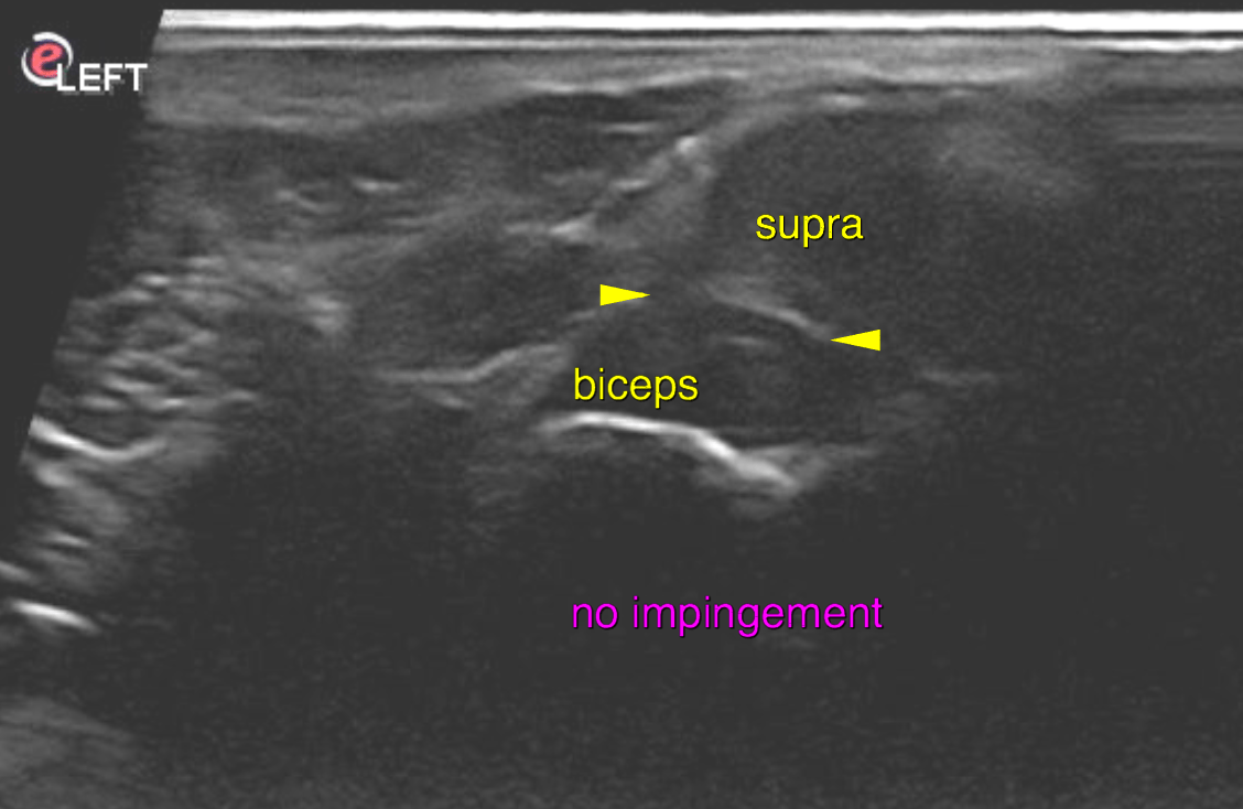
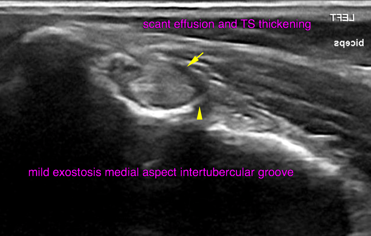
Ultrasound of the left shoulder- Minor effusion and synovial thickening of the left tendon sheath is noted. A mild exostosis formation is seen within the medial and proximal aspect of the inter tubercular groove.
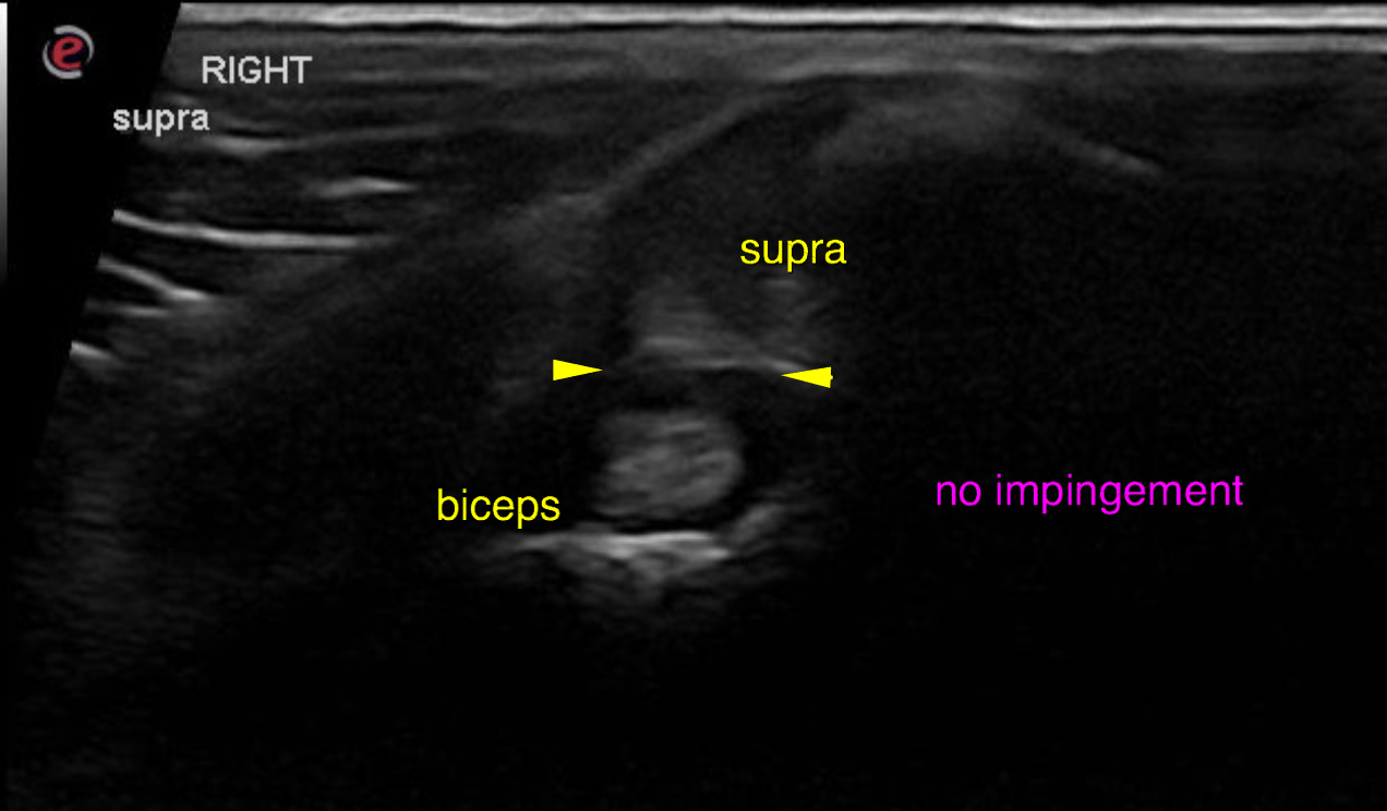
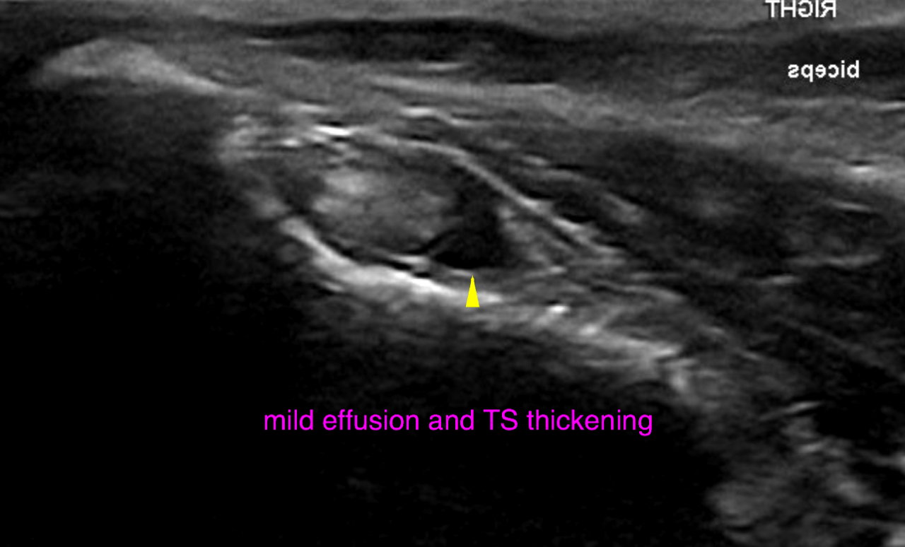
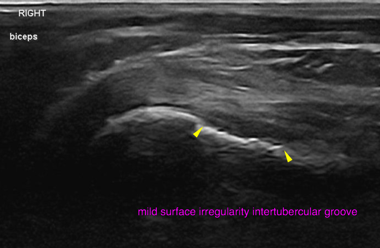
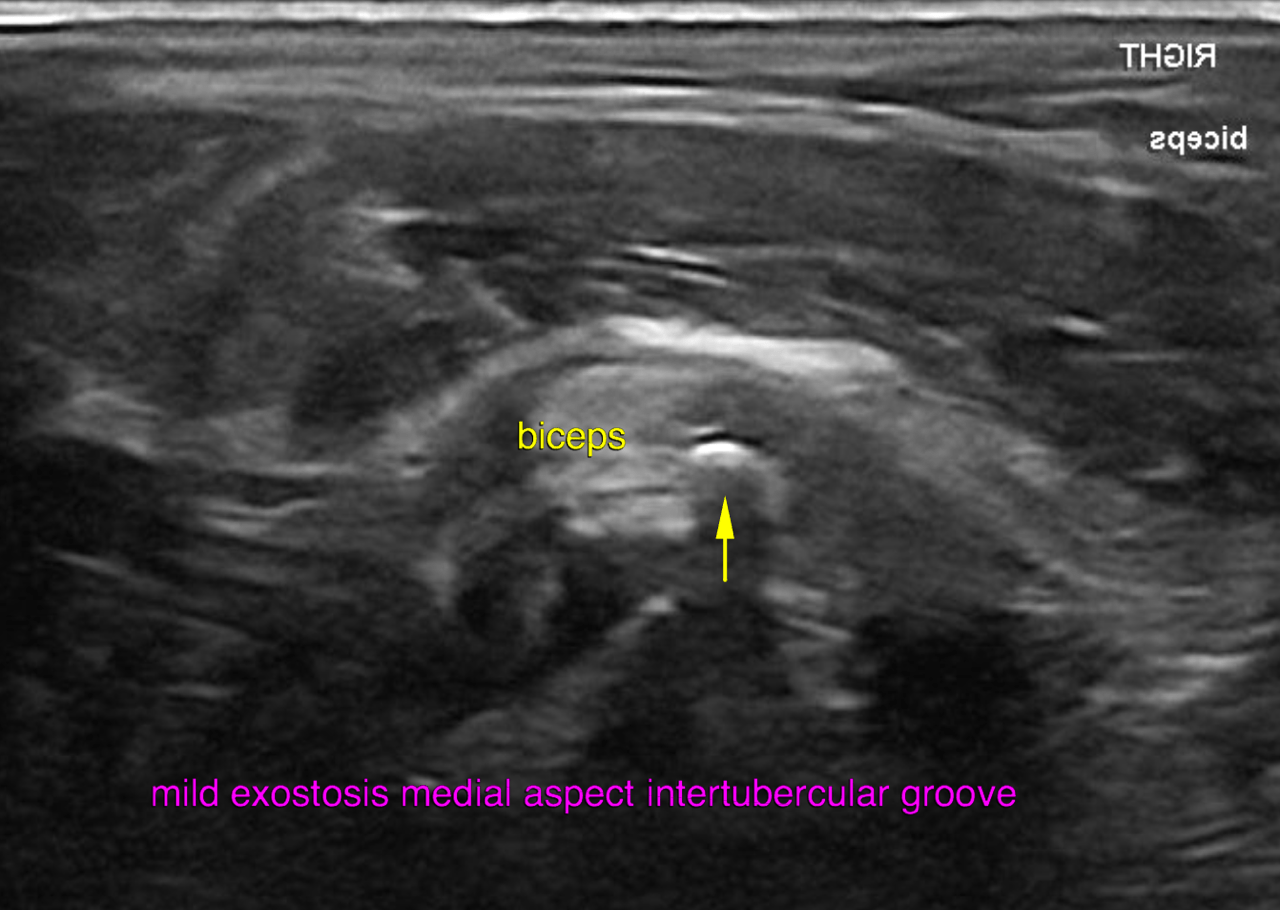
Ultrasound of the right shoulder – Mild effusion and synovial thickening of the tendon sheath is noted. A mild exostosis formation is seen within the medial and proximal aspect of the intertubercular groove.
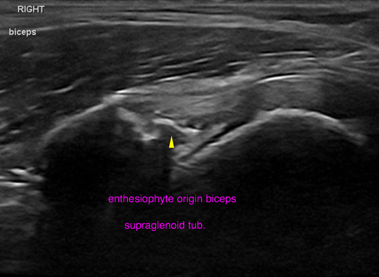
The bone surface of the intertubercular groove is mildly irregular. An enthesiophyte is
associated with the origin of the biceps tendon at the supraglenoid tubercle. The shoulder joint reveals a moderate amount of periarticular osteophyte and a mild
amount of anechoic effusion.

DX

The ultrasonographic findings are compatible with minor chronic bilateral tendovaginitis of the biceps. The changes are slightly more pronounced on the right side.

Outcome

Insertional tendinopathy of the right biceps is noted as well.
Mild osteoarthritis of the right shoulder joint is seen.
The slightly higher degree of tendon sheath effusion on the right side is likely to reflect
joint effusion rather than true tendon sheath effusion.
Overall the findings are very mild and their clinical significance is questionable.

Comments

Based on the radiographs (sent for review as part of the ultrasound but not for an official rad read) there is leftsided medial coronoid pathology (MCP) and bilateral carpal as well as metacarpophalangeal osteoarthritis, which all – but specifically the MCP – are more likely to be the underlying cause of the lameness here. Clinical correlation is warranted and of utmost importance of course. Some minor insertional desmopathy of the interosseus radioulnar ligament/RUIN is noted as well.

Patient Information

Patient Name : Pye Lengi/Skylos
Gender : Male, Neutered
Species : Canine
Type of Imaging : Ultrasound
Status : Complete
Liz Wuz Here : Yes
Code : 18_00002

Clinical Signs

  • Lameness

Images

bildschirmfoto_2016-05-05_um_08bildschirmfoto_2016-05-05_um_08bildschirmfoto_2016-05-05_um_08bildschirmfoto_2016-05-05_um_08bildschirmfoto_2016-05-05_um_08bildschirmfoto_2016-05-05_um_08bildschirmfoto_2016-05-05_um_08bildschirmfoto_2016-05-05_um_08

Clinical Signs

  • Lameness