ORTHO – Bilateral chronic focal tendinitis of the distal insertion tendon of the quadriceps femoris muscle in a 9 year old MN Nova Scotia Duck Retriever dog

Case Study

ORTHO – Bilateral chronic focal tendinitis of the distal insertion tendon of the quadriceps femoris muscle in a 9 year old MN Nova Scotia Duck Retriever dog

This 9 year old MN Nova Scotia Duck Retriever dog presented with progressive left hind limb lameness of 5 months duration. 

Physical exam: grade III/V left pelvic limb lameness with moderate muscle atrophy. Stable left stifle with no palpable effusion, no pain on range of motion. Palpable firm swelling lateral and proximal to patella, moderate pain. 
This 9 year old MN Nova Scotia Duck Retriever dog presented with progressive left hind limb lameness of 5 months duration. 

Physical exam: grade III/V left pelvic limb lameness with moderate muscle atrophy. Stable left stifle with no palpable effusion, no pain on range of motion. Palpable firm swelling lateral and proximal to patella, moderate pain. 
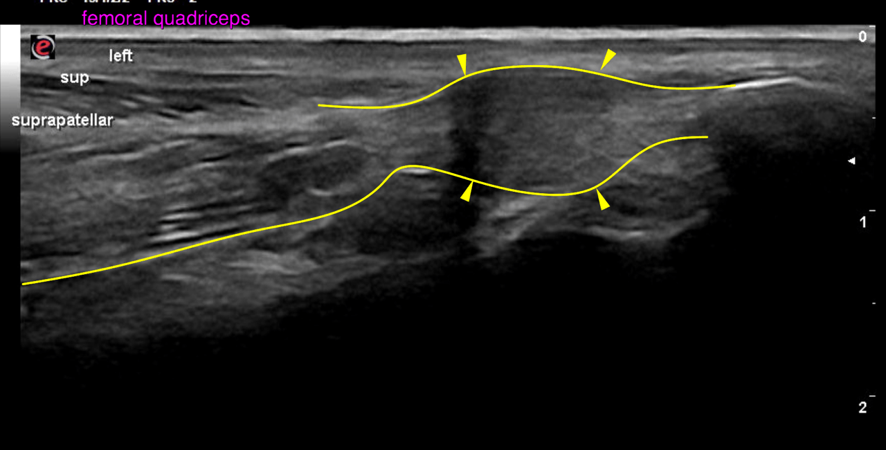
Image Interpretation

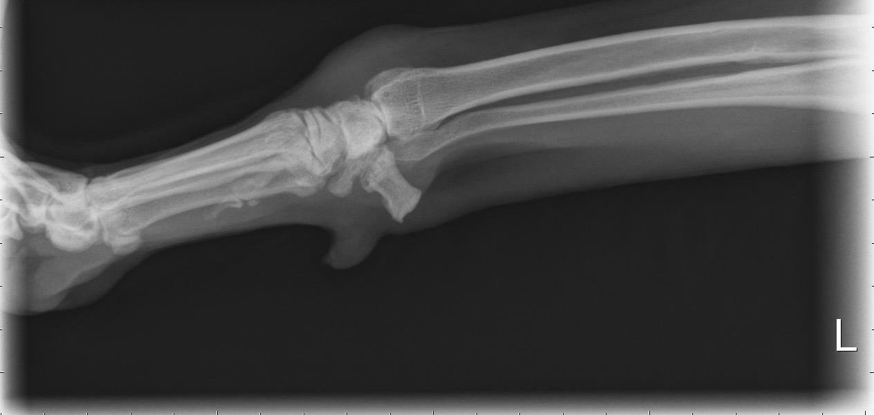
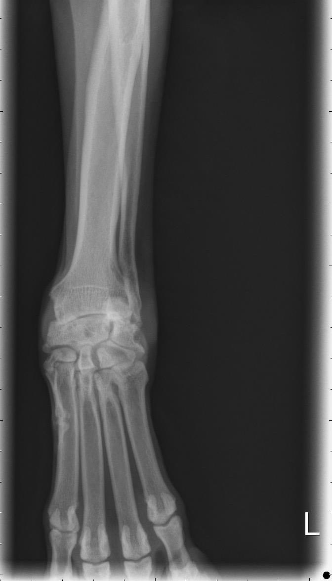
Rads of left carpus: There is a moderate soft tissue swelling circumferential to the left antebrachiocarpal
joint. A mild amount of osteophytes is associated with the periarticular margins of the
left antebrachiocarpal joint. The intercarpal and carpometacarpal joints reveal
emerging osteophytes. An enthesiophyte is seen at the proximopalmar aspect of the
accessory carpal bone.
There is a exostosis-like bony spur at the medial styloid of the radius level with the
sulcus of the abductor pollicis longus muscle.
The intermedioradial carpal bone presents moderate generalized medullary sclerosis
obscuring the trabecular bone pattern and multifocal central cystic lesions.

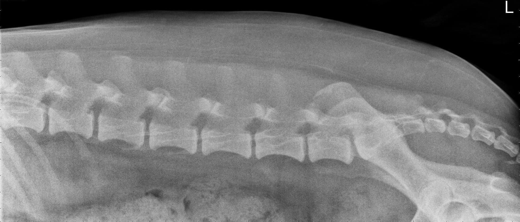
Rads of lumbar spine: The L6/7 disc space presents minor narrowing.

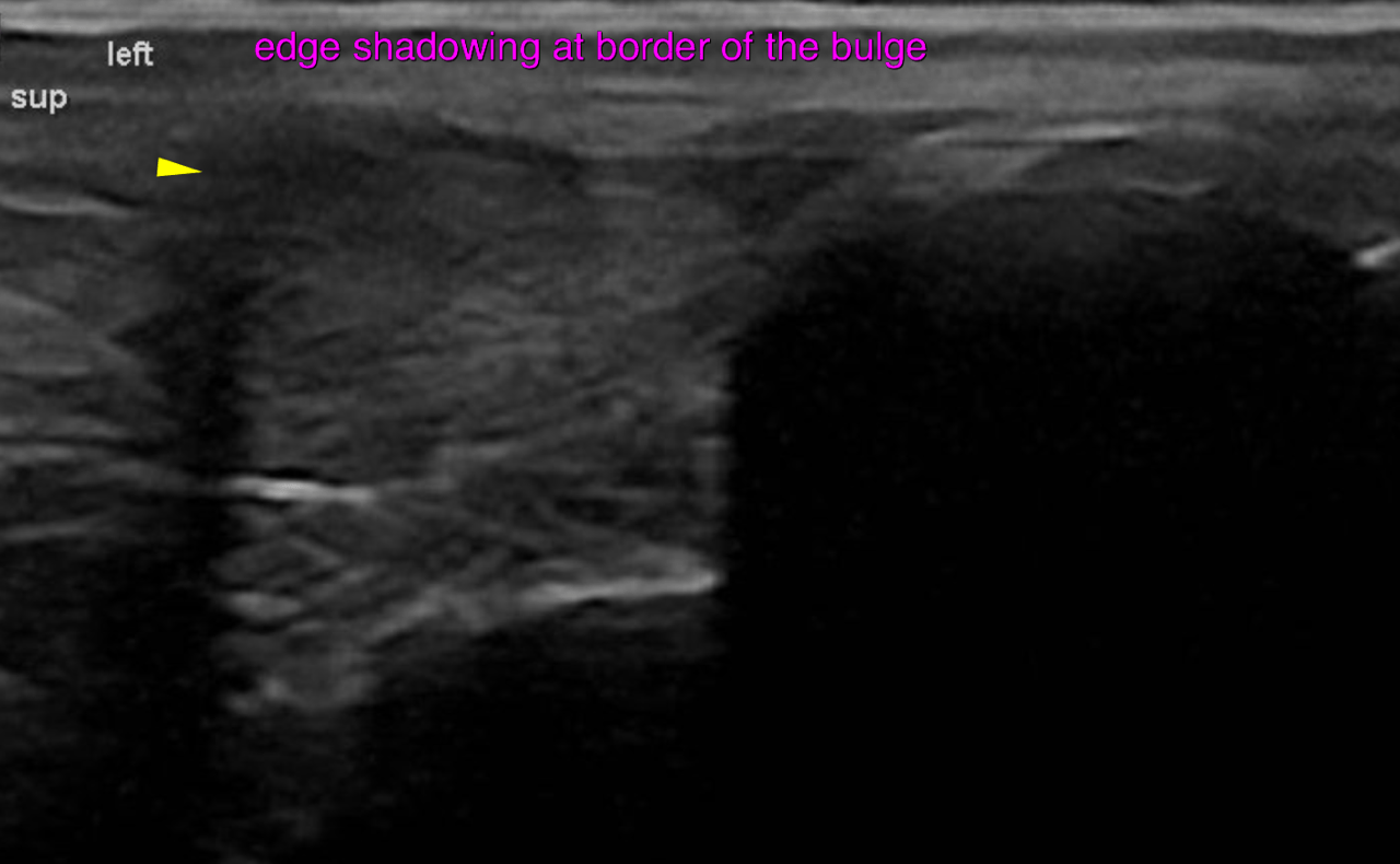
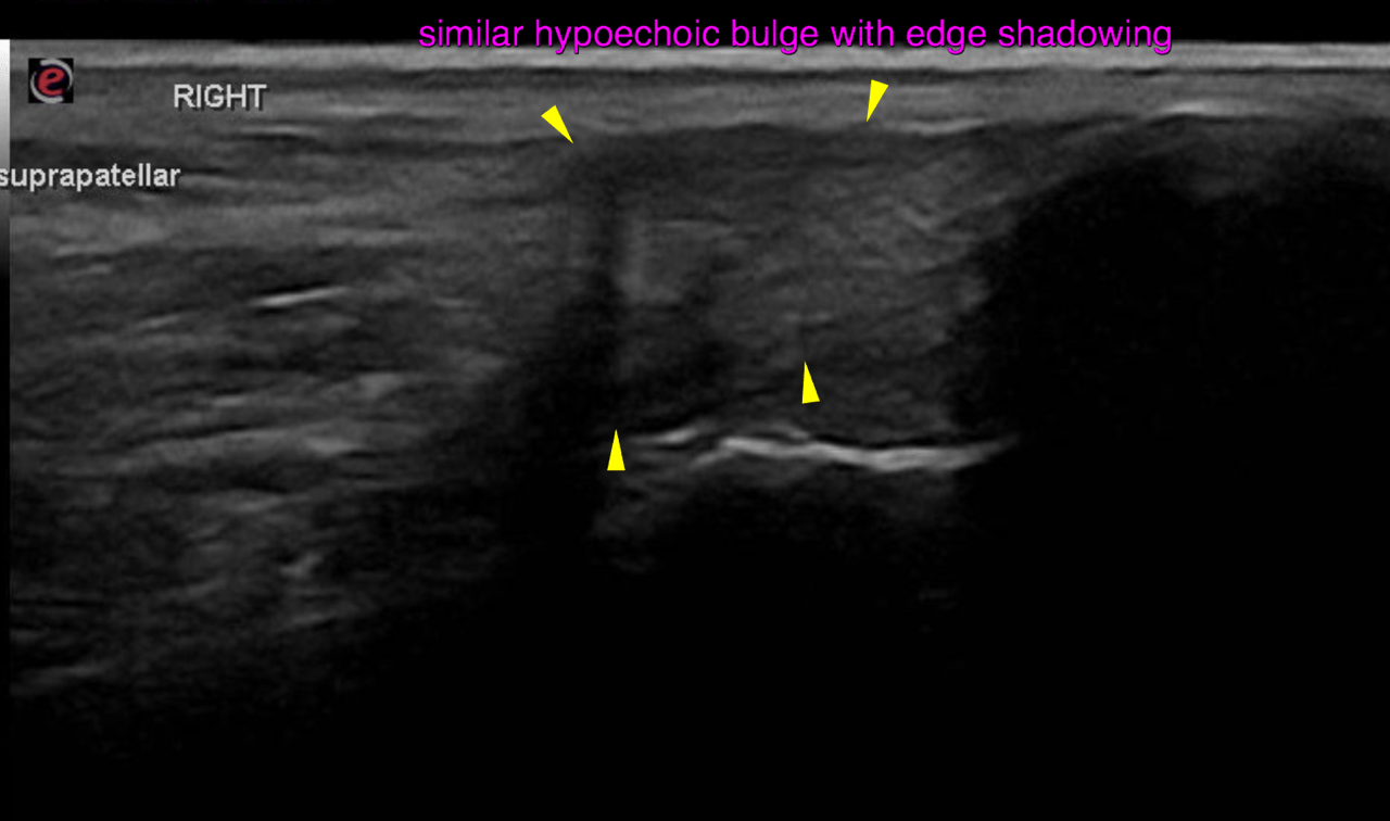
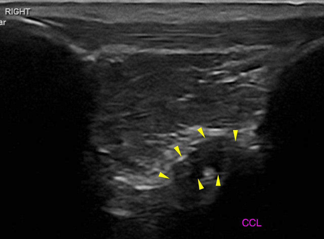
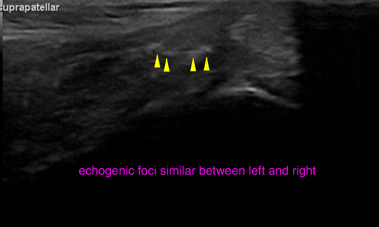
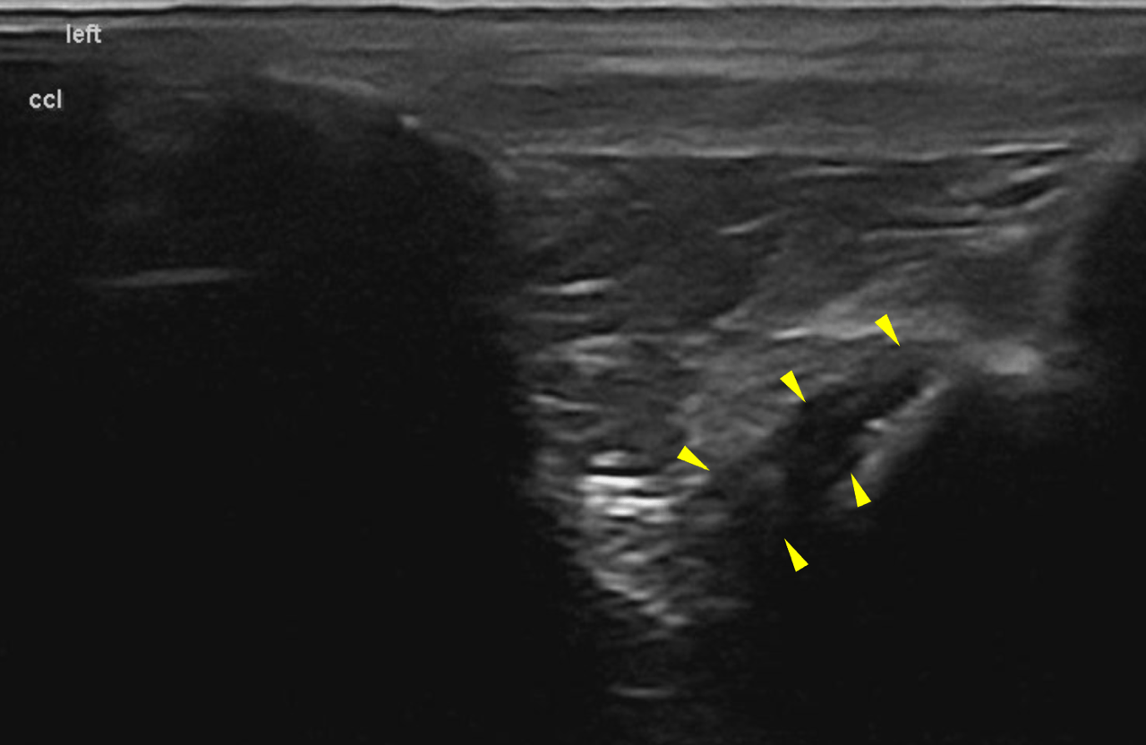
Ultrasound of both stifles reveals the following, less pronounced in the right stifle: focal thickening of the insertion tendon of the quadriceps femoris muscle is seen 5
mm proximal to the base of the patella. The thickened area spans 8 x 7 mm. It reveals a
mild overall decrease in echogenicity with multiple small non-shadowing echogenic
foci. The regular fibre pattern is obscured.
Acoustic shadowing is noted at the most proximal extent of the affected region, which
is a composite effect of edge shadowing & bending reflex as well as mild focal
thickening of the peritendineum.

DX

Bilateral chronic focal tendinitis of the distal insertion tendon of the quadriceps femoris muscle – which is moderate on the left and mild in degree on the right side. • Moderate chronic osteoarthritis of the left antebrachiocarpal joint with indirect signs of abductor pollicis tendovaginitis • Indirect signs of intervertebral disc disease L6/7

Outcome

Focal tendinitis of the quadriceps muscle in this very typical location is usually as an incidental finding. It is assumed to be a result of chronic repetitive microtrauma, abnormal mechanical, degeneration or a combination of these. The findings are relatively pronounced on the left side. This may not be the actual cause of the lameness and muscle atrophy in this
dog. Consider rest, physical therapy as well as local PRP injection as clinical trial. Other
pathology including neurologic has to be considered as well. According to the history the changes in the left carpus do not seem to be of clinical
relevance at this point.

Patient Information

Patient Name : Finley King/Skylos
Gender : Male, Neutered
Species : Canine
Type of Imaging : Ultrasound
Status : Complete
Liz Wuz Here : Yes
Code : 18_00001

Clinical Signs

  • Lameness

Exam Finding

  • Pain

Images

bildschirmfoto_2016-05-06_um_11bildschirmfoto_2016-05-06_um_11bildschirmfoto_2016-05-06_um_13bildschirmfoto_2016-05-06_um_13bildschirmfoto_2016-05-06_um_13bildschirmfoto_2016-05-06_um_13img_3373img_3375img_3377

Clinical Signs

  • Lameness