ORTHO – Chronic degenerative joint disease (DJD) with partial cranial cruciate ligament (CCL) rupture in a 5 year old FS Saint Bernard dog

Case Study

ORTHO – Chronic degenerative joint disease (DJD) with partial cranial cruciate ligament (CCL) rupture in a 5 year old FS Saint Bernard dog

This 5 year old FS Saint Bernard dog presented with left hind acute lameness. Partial weight-bearing.

This 5 year old FS Saint Bernard dog presented with left hind acute lameness. Partial weight-bearing.

Image Interpretation

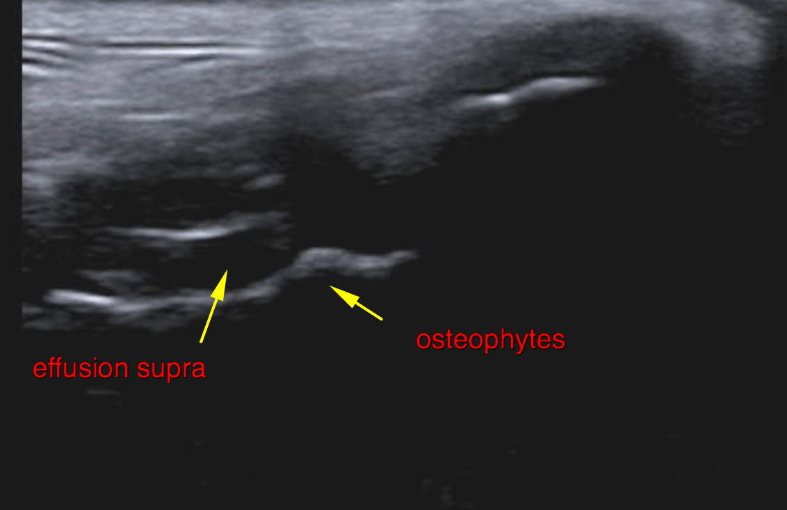
Ultrasound of the left stifle – Moderate anechoic effusion and moderate synovial thickening is noted within the
suprapatellar recess and infrapatellar recess of the right stifle joint.
A moderate amount of osteophytes noted at the periarticular margins of the
femoropatellar & femorotibial joint.
The cranial cruciate ligament presents as a short echogenic stump at the intercondylar
eminence of the tibia. A thin trace of continuous fibres with an irregularly undulating
course is seen.
Marked heterogeneity of the infrapatellar fat pad is noted.
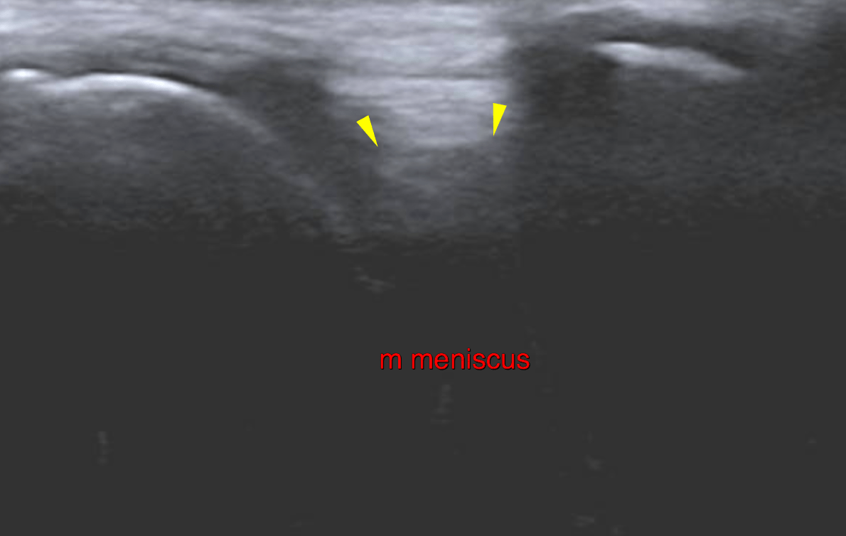
The visible portion of the medial meniscus is in situ, uniform in echogenicity and
smooth in outline. The caudal horn and deep portion of the medial meniscus is not
seen.
DX

The ultrasonographic examination of the left stifle joint is compatible with moderate chronic degenerative joint disease with cranial cruciate ligament failure. The findings indicate a partial rupture with a small portion of maintained fibres. Yet full biomechanical failure is assumed. There is no evidence of meniscal injury at this point.

Patient Information

Patient Name : Daisy Kivela/Bayshore
Gender : Female, Spayed
Species : Canine
Type of Imaging : Ultrasound
Status : Complete
Liz Wuz Here : Yes

Clinical Signs

  • Lameness

Images

bildschirmfoto_2016-04-26_um_17bildschirmfoto_2016-04-26_um_17bildschirmfoto_2016-04-26_um_17

Clinical Signs

  • Lameness