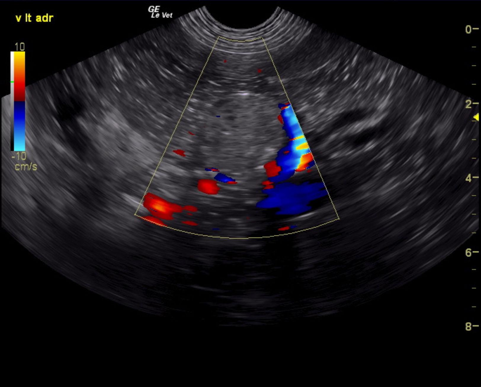
07_00181 CD C Unilateral adrenal mass, +LDDS, nl contralateral adrenal

Case Study

07_00181 CD C Unilateral adrenal mass, +LDDS, nl contralateral adrenal

A 7-year-old SF mixed breed dog was presented for evaluation of PU/PD and polypnea. On physical examination the fur on the flanks was bilaterally thin and she had a pot-bellied appearance. Urinalysis showed SG of 1.023 and 2+ protein. Abnormalities on serum biochemistry were mildly elevated ALT activity (150 U/l). Low-dose dexamethasone suppression showed cortisol of: pre 5.8, 4 hour 5.4; and 8 hour 4.8 mg/dl.


DX

Functional adrenal mass

Sonographic Differential Diagnosis

Left adrenal mass, focal mineralization suggestive for carcinoma. Given the LDDST results functional adenocarcinoma is suspected.

Image Interpretation

Right adrenal uniform 2.55 x 0.62 cm cranial; 0.55 cm caudal. Left adrenal comprised a mass measuring 4 x 2.6 cm caudal; 0.72 cm cranial. Capsular expansion noted without capsular escape. No overt vascular invasion noted. This appears to be resectable. The caudal mass portion of the adrenal measured 2.6 x 0.53 cm deviating the left renal artery caudally.

Outcome

None

Clinical Differential Diagnosis

Pituitary-dependent Cushing’s disease, adrenal adenoma, adrenal carcinoma

Sampling

None

UA Specific Gravity Range

1.023

Patient Information

Gender : Female, Spayed
Species : Canine
Type of Imaging : Ultrasound

Clinical Signs

  • PU-PD
  • Tachypnea

Exam Finding

  • Alopecia
  • Pot belly

Images

cd2cd3cd4cd5

Blood Chemistry

  • ALT (SGPT), High

Clinical Signs

  • PU-PD
  • Tachypnea

Special Testing

  • LDDST Positive for Cushing's

Urinalysi

  • Protein Present
Skip to content