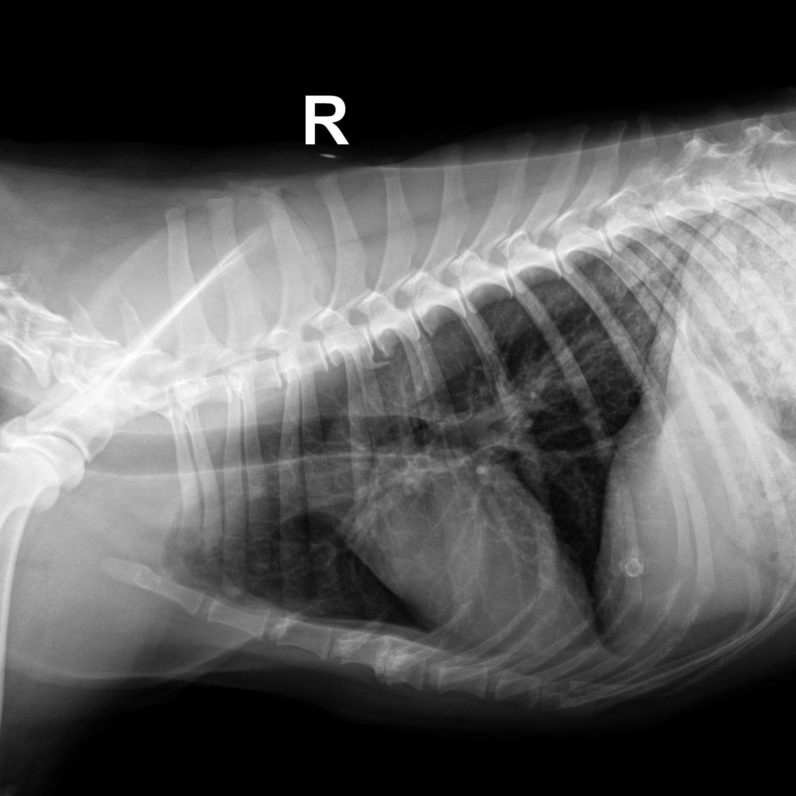
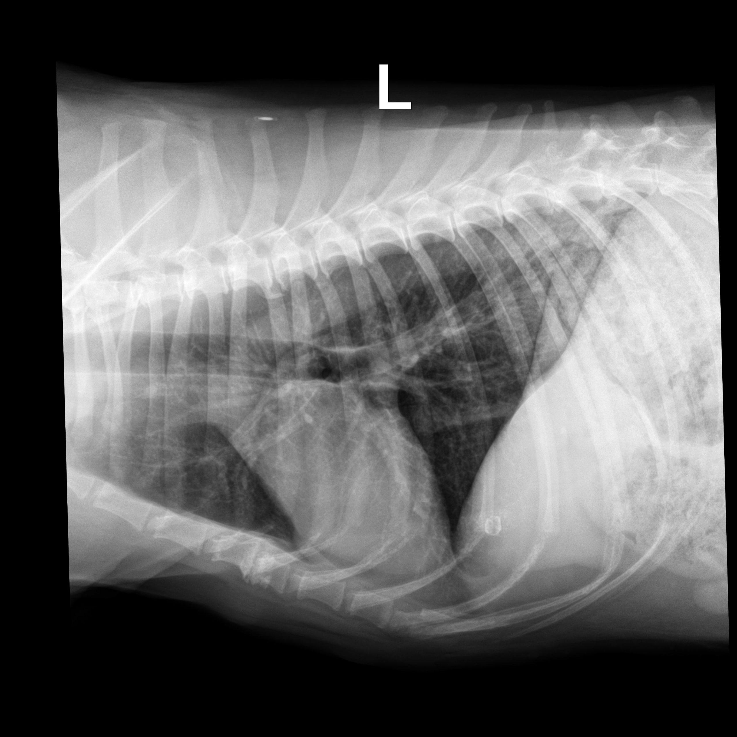
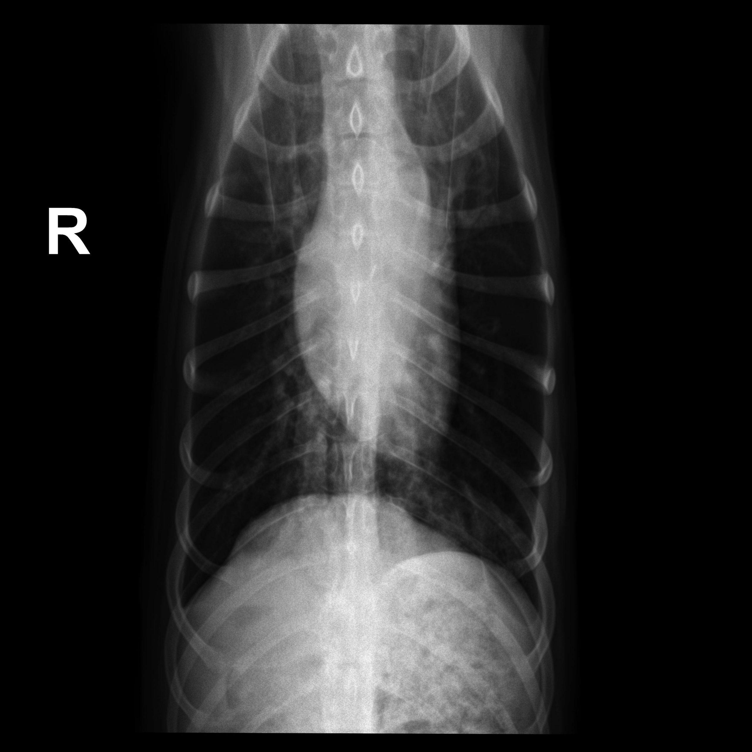
Rads of right lateral, left lateral and VD thorax – Osseous structures: There was mild ventral vertebral endplate modeling within the
mid thoracic spine consistent with spondylosis deformans.
Extrathoracic soft tissue structures: There was a spherical mineral opaque structure
and granulated mineral opaque material superimposed onto the cranial aspect of the
liver and caudal aspect of the lung respectively.
Intrathoracic structures: The course of the trachea was normal. The cardiac silhouette
was small, the caudal vena cava and the pulmonary vasculature were thin.
There was an area of diffusely increased interstitial opacity within the caudodorsal
aspect of the lung.
There also was a generalized bronchial pattern with increased wall visibility and
mineralization.
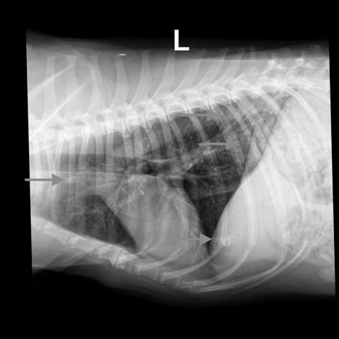
There was a small soft tissue opaque nodule associated with the cranial lobar vessels
best visible on the left lateral view (see image below)


