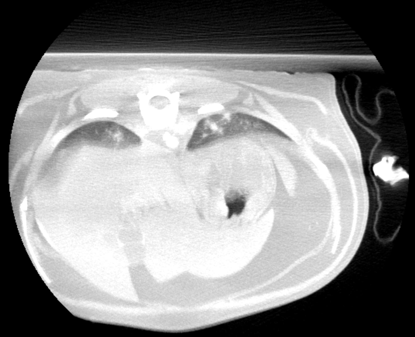
CT of the chest and skull: The chest scan provided covers only the caudal portion of both caudal lung lobes and the cranial abdomen. Within the image aspects of both caudal lung lobes, there are multi focal, soft tissue, attenuating, ill defined interstitial nodules with heterogenous contrast uptake. Atelectatic changes are noted in the dorsal aspects of the lung lobes as the cat was positioned in dorsal recumbency. Within the cranial abdomen, the gallbladder presents bipartite, which is an incidental finding in cats. The pancreas is moderately to severely enlarged and shows multi focal nodular and mass-like lesions expanding the pancreatic capsule. No peripheral fat stranding was noted surrounding the pancreas. The portal lymph nodes both show moderate to severe enlargement, rounding and heterogenous contrast uptake and there are several nodules of varying size within the splenic parenchyma expanding the capsule. Contrast media pooling within the hepatic veins and portal vein, which is likely a function of systemic hypotension under general anesthesia, may also be an indicator of beginning portal hypertension, although no primary changes of the liver were evident from the CT scan. Incidental finding of a mild, emerging spondylosis deformans is noted at multiple sites in the caudal thoracic and cranial lumbar spine. There is a space occupying lesion originating from the mandibular salivary gland on the right side and measuring approximately 2.0 cm in diameter. The lesion shows heterogenous contrast enhancement with predominantly peripheral enhancement and non uniform contrast enhancement in the center of the lesion sparing multiple focal areas. There is moderate enlargement and rounding of the medial retropharyngeal lymph node on the right side with heterogenous contrast uptake. Both tympanic bullae are incompletely filled with soft tissue, attenuating homogenous material. Part of this material on the right side presents with character of a broad based, dorsally attached space-occupying lesion and shows a uniform contrast enhancement. Both tympanic bulla walls show moderate, uniform thickening. There is mild swelling of the nasopharyngeal soft tissues, medial to both tympanic bullae. Bilateral chronic otitis media with poly formation on the presumed right side.