CT Malignant Osteolytic Lesion of the Right Zygomatic Arch in a 7 year old Great Dane dog

Case Study

CT Malignant Osteolytic Lesion of the Right Zygomatic Arch in a 7 year old Great Dane dog

The patient was diagnosed with osteosarcoma of the left distal radius 6 months ago. Currently stable on multimodal pain management. Non weight-bearing left front leg, and mass has grown in size. There is acute swelling of the TMJ area (in the last 24 hours). The patient is painful and unwilling to open the mouth to take pain medications or eat. History of partial CCL tear of right hind leg; the dog does wear a stifle orthotic for stability.

The patient was diagnosed with osteosarcoma of the left distal radius 6 months ago. Currently stable on multimodal pain management. Non weight-bearing left front leg, and mass has grown in size. There is acute swelling of the TMJ area (in the last 24 hours). The patient is painful and unwilling to open the mouth to take pain medications or eat. History of partial CCL tear of right hind leg; the dog does wear a stifle orthotic for stability.

Physical exam results: ocular exam WNL, aural exam WNL. There is a soft non-pitting swelling of the right temporal/mandibular region which is sensitive to the touch but no heat appreciated. The patient is reluctant to have the mouth opened. A brief oral exam yielded NSF. Heart and lungs auscultate WNL. No overt neurological deficits. Non-weight bearing lameness of left front leg. 

Body weight 102#, Temp 102.5, Pulse 80, respiratory rate 20, mucous membranes pink, CRT 2 seconds.CBC is WNL, Chemistry profile slightly low T4 and TSH is low end normal.
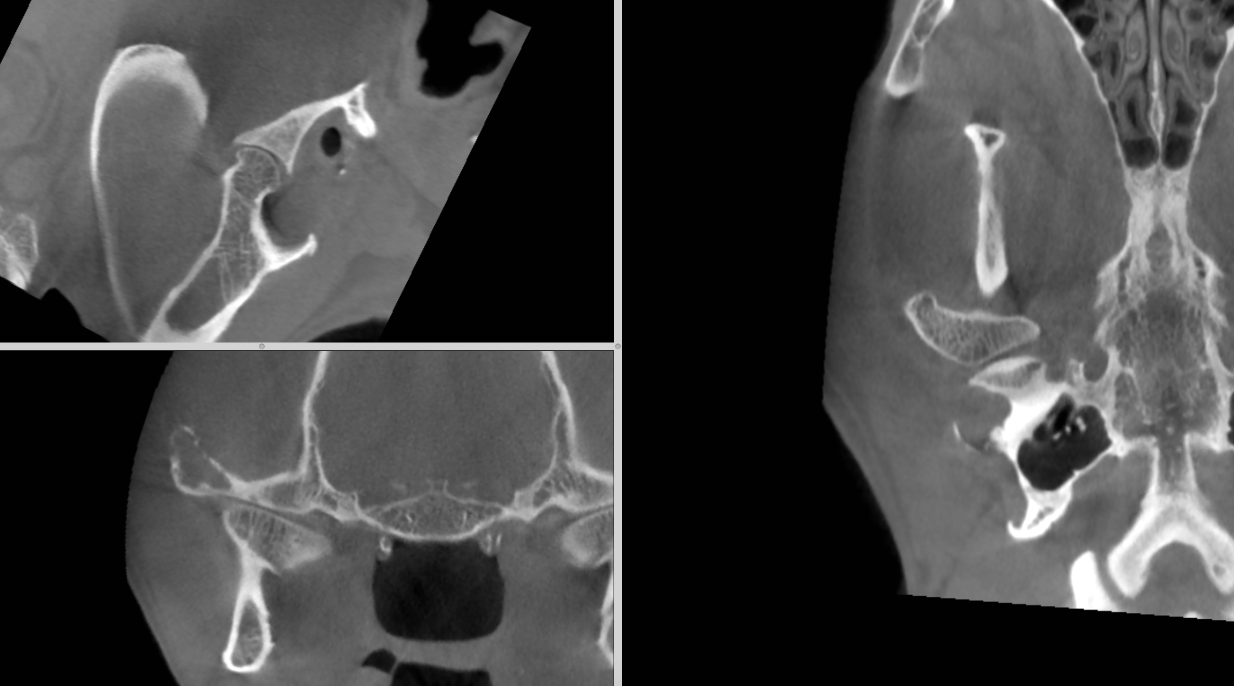
DX

High resolution CT of the head revealed a monostotic highly aggressive osteolytic lesion of the right zygomatic arch adjacent to the TMJ. The CT signs clearly indicate malignancy. Considering the history of the patient metastatic spread of the prediagnosed radial osteosarcoma is a very high potential.

Image Interpretation

There was a mild soft tissue swelling noted lateral to the right temporomandibular joint (TMJ). A permeative osteolytic region with a long transition zone to the normal bone was seen within the zygomatic process of the right temporal bone extending to the mid portion of the zygomatic arch. Significant cortical thinning and multifocal interruptions of the bony cortex were present. Amorphous and sunburst-like periosteal reactions were present onto the bone surface. The lesion was not crossing the joint. No osteolytic defects were seen within the adjacent mandibular fossa and the condylar process itself. The TM joint space presented mild generalized widening as compared with the left TMJ.
The ipsilateral medial retropharyngeal lymph node presented no abnormalities. The submandibular nodes were not included in the study.

Outcome

The prognosis is poor. For final assessment of prognosis and discussion of a potential (palliative) treatment plan full oncological staging would be required.

Patient Information

Patient Name : Lucy Cormier, Into the Sunset
Gender : Female, Spayed
Species : Canine
Type of Imaging : Ultrasound
Status : Complete

Clinical Signs

  • Lameness
  • Palpable Mass

History

  • Neoplasia

Exam Finding

  • Palpable mass

Images

bildschirmfoto_2015-06-24_um_10

Blood Chemistry

  • Hypothyroidism

Clinical Signs

  • Lameness
  • Palpable Mass
Skip to content